 Movie
Movie Controller


+ Open data

- Basic information
| Entry | Database: PDB / ID: 8wey | ||||||
|---|---|---|---|---|---|---|---|
| Title | PSI-LHCI of the red alga Cyanidium caldarium RK-1 (NIES-2137) | ||||||
 Components Components |
| ||||||
 Keywords Keywords | PHOTOSYNTHESIS / Photosystem I / ELECTRON TRANSPORT | ||||||
| Function / homology | : / BETA-CAROTENE / CHLOROPHYLL A ISOMER / CHLOROPHYLL A / DIGALACTOSYL DIACYL GLYCEROL (DGDG) / 1,2-DIPALMITOYL-PHOSPHATIDYL-GLYCEROLE / PHYLLOQUINONE / IRON/SULFUR CLUSTER / Unknown ligand Function and homology information Function and homology information | ||||||
| Biological species |  Cyanidium caldarium (eukaryote) Cyanidium caldarium (eukaryote) | ||||||
| Method | ELECTRON MICROSCOPY / single particle reconstruction / cryo EM / Resolution: 1.92 Å | ||||||
 Authors Authors | Kato, K. / Hamaguchi, T. / Nakajima, Y. / Kawakami, K. / Yonekura, K. / Shen, J.R. / Nagao, R. | ||||||
| Funding support | 1items
| ||||||
 Citation Citation | Journal: Proc Natl Acad Sci U S A / Year: 2024 Title: The structure of PSI-LHCI from provides evolutionary insights into conservation and diversity of red-lineage LHCs. Authors: Koji Kato / Tasuku Hamaguchi / Minoru Kumazawa / Yoshiki Nakajima / Kentaro Ifuku / Shunsuke Hirooka / Yuu Hirose / Shin-Ya Miyagishima / Takehiro Suzuki / Keisuke Kawakami / Naoshi Dohmae / ...Authors: Koji Kato / Tasuku Hamaguchi / Minoru Kumazawa / Yoshiki Nakajima / Kentaro Ifuku / Shunsuke Hirooka / Yuu Hirose / Shin-Ya Miyagishima / Takehiro Suzuki / Keisuke Kawakami / Naoshi Dohmae / Koji Yonekura / Jian-Ren Shen / Ryo Nagao /  Abstract: Light-harvesting complexes (LHCs) are diversified among photosynthetic organisms, and the structure of the photosystem I-LHC (PSI-LHCI) supercomplex has been shown to be variable depending on the ...Light-harvesting complexes (LHCs) are diversified among photosynthetic organisms, and the structure of the photosystem I-LHC (PSI-LHCI) supercomplex has been shown to be variable depending on the species of organisms. However, the structural and evolutionary correlations of red-lineage LHCs are unknown. Here, we determined a 1.92-Å resolution cryoelectron microscopic structure of a PSI-LHCI supercomplex isolated from the red alga RK-1 (NIES-2137), which is an important taxon in the Cyanidiophyceae. We subsequently investigated the correlations of PSI-LHCIs from different organisms through structural comparisons and phylogenetic analysis. The PSI-LHCI structure obtained shows five LHCI subunits surrounding a PSI-monomer core. The five LHCIs are composed of two Lhcr1s, two Lhcr2s, and one Lhcr3. Phylogenetic analysis of LHCs bound to PSI in the red-lineage algae showed clear orthology of LHCs between and , whereas no orthologous relationships were found between Lhcr1-3 and LHCs in other red-lineage PSI-LHCI structures. These findings provide evolutionary insights into conservation and diversity of red-lineage LHCs associated with PSI. | ||||||
| History |
|
- Structure visualization
| Structure viewer | Molecule: MolmilJmol/JSmol |
|---|
- Downloads & links
-Download
| PDBx/mmCIF format | 8wey.cif.gz | 908.5 KB | Display | PDBx/mmCIF format |
|---|---|---|---|---|
| PDB format | pdb8wey.ent.gz | 792.9 KB | Display | PDB format |
| PDBx/mmJSON format | 8wey.json.gz | Tree view | PDBx/mmJSON format | |
| Others |  Other downloads Other downloads |
-Validation report
| Arichive directory | https://data.pdbj.org/pub/pdb/validation_reports/we/8weyftp://data.pdbj.org/pub/pdb/validation_reports/we/8wey | HTTPS FTP |
|---|
-Related structure data
| Related structure data |  37480MC M: map data used to model this data C: citing same article ( |
|---|---|
| Similar structure data |
-Links
 PDBj
PDBj


- Assembly
| Deposited unit | 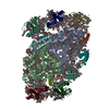
|
|---|---|
| 1 |
|
-Components
-Photosystem I ... , 12 types, 12 molecules ABCDEFIJKLMO
| #1: Protein | Mass: 82698.344 Da / Num. of mol.: 1 / Source method: isolated from a natural source / Source: (natural) Cyanidium caldarium (eukaryote) |
|---|---|
| #2: Protein | Mass: 82160.898 Da / Num. of mol.: 1 / Source method: isolated from a natural source / Source: (natural) Cyanidium caldarium (eukaryote) |
| #3: Protein | Mass: 8822.272 Da / Num. of mol.: 1 / Source method: isolated from a natural source / Source: (natural) Cyanidium caldarium (eukaryote) |
| #4: Protein | Mass: 15709.957 Da / Num. of mol.: 1 / Source method: isolated from a natural source / Source: (natural) Cyanidium caldarium (eukaryote) |
| #5: Protein | Mass: 6974.055 Da / Num. of mol.: 1 / Source method: isolated from a natural source / Source: (natural) Cyanidium caldarium (eukaryote) |
| #6: Protein | Mass: 20520.506 Da / Num. of mol.: 1 / Source method: isolated from a natural source / Source: (natural) Cyanidium caldarium (eukaryote) |
| #7: Protein/peptide | Mass: 3408.183 Da / Num. of mol.: 1 / Source method: isolated from a natural source / Source: (natural) Cyanidium caldarium (eukaryote) |
| #8: Protein/peptide | Mass: 4426.245 Da / Num. of mol.: 1 / Source method: isolated from a natural source / Source: (natural) Cyanidium caldarium (eukaryote) |
| #9: Protein | Mass: 7105.433 Da / Num. of mol.: 1 / Source method: isolated from a natural source / Source: (natural) Cyanidium caldarium (eukaryote) |
| #10: Protein | Mass: 15343.729 Da / Num. of mol.: 1 / Source method: isolated from a natural source / Source: (natural) Cyanidium caldarium (eukaryote) |
| #11: Protein/peptide | Mass: 3010.698 Da / Num. of mol.: 1 / Source method: isolated from a natural source / Source: (natural) Cyanidium caldarium (eukaryote) |
| #12: Protein | Mass: 16661.221 Da / Num. of mol.: 1 / Source method: isolated from a natural source / Source: (natural) Cyanidium caldarium (eukaryote) |
-Protein , 3 types, 5 molecules 14253
| #13: Protein | Mass: 24117.104 Da / Num. of mol.: 2 / Source method: isolated from a natural source / Source: (natural) Cyanidium caldarium (eukaryote)#14: Protein | Mass: 24128.066 Da / Num. of mol.: 2 / Source method: isolated from a natural source / Source: (natural) Cyanidium caldarium (eukaryote)#15: Protein | | Mass: 23826.514 Da / Num. of mol.: 1 / Source method: isolated from a natural source / Source: (natural) Cyanidium caldarium (eukaryote) |
|---|
-Sugars , 2 types, 8 molecules 

| #22: Sugar | ChemComp-LMT /  Type: D-saccharide / Mass: 510.615 Da / Num. of mol.: 7 / Source method: obtained synthetically / Formula: C24H46O11 / Comment: detergent*YM Type: D-saccharide / Mass: 510.615 Da / Num. of mol.: 7 / Source method: obtained synthetically / Formula: C24H46O11 / Comment: detergent*YM#24: Sugar | ChemComp-DGD / |  Type: saccharide / Mass: 949.299 Da / Num. of mol.: 1 / Source method: obtained synthetically / Formula: C51H96O15 Type: saccharide / Mass: 949.299 Da / Num. of mol.: 1 / Source method: obtained synthetically / Formula: C51H96O15 |
|---|
-Non-polymers , 9 types, 790 molecules 


| #16: Chemical | ChemComp-CL0 /  Mass: 893.489 Da / Num. of mol.: 1 / Source method: obtained synthetically / Formula: C55H72MgN4O5 Mass: 893.489 Da / Num. of mol.: 1 / Source method: obtained synthetically / Formula: C55H72MgN4O5 | ||||||||||||||
|---|---|---|---|---|---|---|---|---|---|---|---|---|---|---|---|
| #17: Chemical | ChemComp-CLA /  Mass: 893.489 Da / Num. of mol.: 153 / Source method: obtained synthetically / Formula: C55H72MgN4O5 Mass: 893.489 Da / Num. of mol.: 153 / Source method: obtained synthetically / Formula: C55H72MgN4O5#18: Chemical |  Mass: 450.696 Da / Num. of mol.: 2 / Source method: obtained synthetically / Formula: C31H46O2 Mass: 450.696 Da / Num. of mol.: 2 / Source method: obtained synthetically / Formula: C31H46O2#19: Chemical |  Mass: 722.970 Da / Num. of mol.: 3 / Source method: obtained synthetically / Formula: C38H75O10P / Comment: phospholipid*YM Mass: 722.970 Da / Num. of mol.: 3 / Source method: obtained synthetically / Formula: C38H75O10P / Comment: phospholipid*YM#20: Chemical | ChemComp-BCR /  Mass: 536.873 Da / Num. of mol.: 20 / Source method: obtained synthetically / Formula: C40H56 Mass: 536.873 Da / Num. of mol.: 20 / Source method: obtained synthetically / Formula: C40H56#21: Chemical |  Mass: 351.640 Da / Num. of mol.: 3 / Source method: obtained synthetically / Formula: Fe4S4 Mass: 351.640 Da / Num. of mol.: 3 / Source method: obtained synthetically / Formula: Fe4S4#23: Chemical | ChemComp-UNL / Num. of mol.: 25 / Source method: obtained synthetically #25: Chemical | ChemComp-5X6 / Mass: 568.871 Da / Num. of mol.: 23 / Source method: obtained synthetically / Formula: C40H56O2 #26: Water | ChemComp-HOH / | Mass: 18.015 Da / Num. of mol.: 560 / Source method: isolated from a natural source / Formula: H2O |
-Details
| Has ligand of interest | N |
|---|---|
| Has protein modification | Y |
-Experimental details
-Experiment
| Experiment | Method: ELECTRON MICROSCOPY |
|---|---|
| EM experiment | Aggregation state: PARTICLE / 3D reconstruction method: single particle reconstruction |
- Sample preparation
| Component | Name: PSI-LHCI / Type: COMPLEX / Entity ID: #1-#15 / Source: NATURAL | |||||||||||||||
|---|---|---|---|---|---|---|---|---|---|---|---|---|---|---|---|---|
| Molecular weight | Value: 0.55 MDa / Experimental value: NO | |||||||||||||||
| Source (natural) | Organism: Cyanidium caldarium (eukaryote) | |||||||||||||||
| Buffer solution | pH: 6.5 | |||||||||||||||
| Buffer component |
| |||||||||||||||
| Specimen | Conc.: 2.83 mg/ml / Embedding applied: NO / Shadowing applied: NO / Staining applied: NO / Vitrification applied: YES | |||||||||||||||
| Specimen support | Grid mesh size: 200 divisions/in. / Grid type: Quantifoil R0.6/1 | |||||||||||||||
| Vitrification | Instrument: FEI VITROBOT MARK IV / Cryogen name: ETHANE / Humidity: 100 % / Chamber temperature: 277 K |
- Electron microscopy imaging
| Microscopy | Model: JEOL CRYO ARM 300 |
|---|---|
| Electron gun | Electron source:  FIELD EMISSION GUN / Accelerating voltage: 300 kV / Illumination mode: FLOOD BEAM FIELD EMISSION GUN / Accelerating voltage: 300 kV / Illumination mode: FLOOD BEAM |
| Electron lens | Mode: BRIGHT FIELD / Nominal defocus max: 1800 nm / Nominal defocus min: 800 nm |
| Image recording | Electron dose: 39.8 e/Å2 / Detector mode: COUNTING / Film or detector model: GATAN K3 (6k x 4k) |
- Processing
| EM software |
| ||||||||||||||||||||||||||||||||||||
|---|---|---|---|---|---|---|---|---|---|---|---|---|---|---|---|---|---|---|---|---|---|---|---|---|---|---|---|---|---|---|---|---|---|---|---|---|---|
| CTF correction | Type: PHASE FLIPPING AND AMPLITUDE CORRECTION | ||||||||||||||||||||||||||||||||||||
| 3D reconstruction | Resolution: 1.92 Å / Resolution method: FSC 0.143 CUT-OFF / Num. of particles: 228449 / Symmetry type: POINT | ||||||||||||||||||||||||||||||||||||
| Atomic model building | Protocol: FLEXIBLE FIT / Space: RECIPROCAL | ||||||||||||||||||||||||||||||||||||
| Atomic model building | Details: homology model / Source name: Other / Type: in silico model |
