Japan Agency for Medical Research and Development (AMED)
Japan
Citation
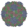
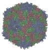
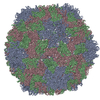
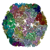
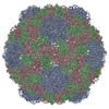
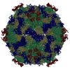
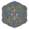
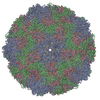
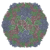
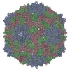
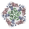
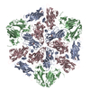
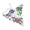
Journal: Commun Biol / Year: 2020 Title: Apple latent spherical virus structure with stable capsid frame supports quasi-stable protrusions expediting genome release. Authors: Hisashi Naitow / Tasuku Hamaguchi / Saori Maki-Yonekura / Masamichi Isogai / Nobuyuki Yoshikawa / Koji Yonekura / Abstract: Picorna-like plant viruses are non-enveloped RNA spherical viruses of ~30 nm. Part of the survival of these viruses depends on their capsid being stable enough to harbour the viral genome and yet ...Picorna-like plant viruses are non-enveloped RNA spherical viruses of ~30 nm. Part of the survival of these viruses depends on their capsid being stable enough to harbour the viral genome and yet malleable enough to allow its release. However, molecular mechanisms remain obscure. Here, we report a structure of a picorna-like plant virus, apple latent spherical virus, at 2.87 Å resolution by single-particle cryo-electron microscopy (cryo-EM) with a cold-field emission beam. The cryo-EM map reveals a unique structure composed of three capsid proteins Vp25, Vp20, and Vp24. Strikingly Vp25 has a long N-terminal extension, which substantially stabilises the capsid frame of Vp25 and Vp20 subunits. Cryo-EM images also resolve RNA genome leaking from a pentameric protrusion of Vp24 subunits. The structures and observations suggest that genome release occurs through occasional opening of the Vp24 subunits, possibly suppressed to a low frequency by the rigid frame of the other subunits.
Idetical with deposited unit in distinct coordinate
icosahedral asymmetric unit, std point frame
Type
Name
Symmetry operation
Number
transform to point frame
1
Symmetry
Point symmetry: (Schoenflies symbol: I (icosahedral))
-
Components
#1: Protein
VP20protein
Mass: 19957.994 Da / Num. of mol.: 1 / Source method: isolated from a natural source / Source: (natural) Apple latent spherical virus / References: UniProt: Q9JGP1
#2: Protein
VP24protein
Mass: 21575.467 Da / Num. of mol.: 1 / Source method: isolated from a natural source / Source: (natural) Apple latent spherical virus / References: UniProt: Q9JGP1
#3: Protein
VP25protein
Mass: 24098.459 Da / Num. of mol.: 1 / Source method: isolated from a natural source / Source: (natural) Apple latent spherical virus / References: UniProt: Q9JGP1
-
Experimental details
-
Experiment
Experiment
Method: ELECTRON MICROSCOPY
EM experiment
Aggregation state: PARTICLE / 3D reconstruction method: single particle reconstruction
-
Sample preparation
Component
Name: Apple latent spherical virus / Type: VIRUS Details: ALSV was purified and isolated from infected Chenopodium quinoa leaf. Entity ID: all / Source: NATURAL
Molecular weight
Experimental value: NO
Source (natural)
Organism: Apple latent spherical virus
Details of virus
Empty: NO / Enveloped: NO / Isolate: SPECIES / Type: VIRION
Natural host
Organism: Malus domestica
Virus shell
Diameter: 300 nm / Triangulation number (T number): 3
Buffer solution
pH: 7.8
Buffer component
ID
Conc.
Formula
Buffer-ID
1
0.1M
Tris-HCl
1
2
0.1M
NaCl
1
3
5mM
MgCl
1
Specimen
Conc.: 1 mg/ml / Embedding applied: YES / Shadowing applied: NO / Staining applied: NO / Vitrification applied: YES Details: In practice, the sample concentration is 1-2 mg/ml.
Specimen support
Grid material: COPPER / Grid type: Quantifoil
EM embedding
Material: ice
Vitrification
Cryogen name: ETHANE
-
Electron microscopy imaging
Microscopy
Model: JEOL CRYO ARM 300
Electron gun
Electron source: FIELD EMISSION GUN / Accelerating voltage: 300 kV / Illumination mode: FLOOD BEAM
Electron lens
Mode: BRIGHT FIELD / Nominal magnification: 40000 X / Nominal defocus max: 2000 nm / Nominal defocus min: 500 nm
Specimen holder
Cryogen: NITROGEN / Specimen holder model: JEOL
Image recording
Electron dose: 8.5 e/Å2 / Detector mode: SUPER-RESOLUTION / Film or detector model: GATAN K2 SUMMIT (4k x 4k)
-
Processing
Software
Name
Version
Classification
NB
phenix.real_space_refine
1.16_3549
refinement
PHENIX
1.16_3549
refinement
EM software
ID
Name
Category
7
Coot
modelfitting
9
PHENIX
modelrefinement
CTF correction
Type: PHASE FLIPPING AND AMPLITUDE CORRECTION
3D reconstruction
Resolution: 2.87 Å / Resolution method: FSC 0.143 CUT-OFF / Num. of particles: 8018 / Symmetry type: POINT
In the structure databanks used in Yorodumi, some data are registered as the other names, "COVID-19 virus" and "2019-nCoV". Here are the details of the virus and the list of structure data.
Jan 31, 2019. EMDB accession codes are about to change! (news from PDBe EMDB page)
EMDB accession codes are about to change! (news from PDBe EMDB page)
The allocation of 4 digits for EMDB accession codes will soon come to an end. Whilst these codes will remain in use, new EMDB accession codes will include an additional digit and will expand incrementally as the available range of codes is exhausted. The current 4-digit format prefixed with “EMD-” (i.e. EMD-XXXX) will advance to a 5-digit format (i.e. EMD-XXXXX), and so on. It is currently estimated that the 4-digit codes will be depleted around Spring 2019, at which point the 5-digit format will come into force.
The EM Navigator/Yorodumi systems omit the EMD- prefix.
Related info.:Q: What is EMD? / ID/Accession-code notation in Yorodumi/EM Navigator
Yorodumi is a browser for structure data from EMDB, PDB, SASBDB, etc.
This page is also the successor to EM Navigator detail page, and also detail information page/front-end page for Omokage search.
The word "yorodu" (or yorozu) is an old Japanese word meaning "ten thousand". "mi" (miru) is to see.
Related info.:EMDB / PDB / SASBDB / Comparison of 3 databanks / Yorodumi Search / Aug 31, 2016. New EM Navigator & Yorodumi / Yorodumi Papers / Jmol/JSmol / Function and homology information / Changes in new EM Navigator and Yorodumi
 Movie
Movie Controller


 Open data
 Basic information
 Components
 Keywords
 Function and homology information
 Apple latent spherical virus
 Authors
 Japan, 3items
  Citation
 Structure visualization
 Downloads & links
 Other downloads
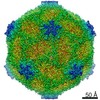
 PDBj
PDBj Assembly

 Sample preparation
 Electron microscopy imaging
 FIELD EMISSION GUN / Accelerating voltage: 300 kV / Illumination mode: FLOOD BEAM
 Processing