Movie
Movie Controller


+ Open data

- Basic information
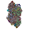
| Entry | Database: PDB / ID: 7d1t | |||||||||||||||||||||||||||||||||||||||||||||
|---|---|---|---|---|---|---|---|---|---|---|---|---|---|---|---|---|---|---|---|---|---|---|---|---|---|---|---|---|---|---|---|---|---|---|---|---|---|---|---|---|---|---|---|---|---|---|
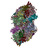
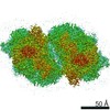
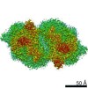
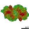
| Title | Cryo-EM Structure of PSII at 1.95 angstrom resolution | |||||||||||||||||||||||||||||||||||||||||||||
 Components Components |
| |||||||||||||||||||||||||||||||||||||||||||||
 Keywords Keywords | PHOTOSYNTHESIS / Photosystem II | |||||||||||||||||||||||||||||||||||||||||||||
| Function / homology |  Function and homology information Function and homology informationphotosystem II oxygen evolving complex / photosystem II assembly / oxygen evolving activity / photosystem II stabilization / photosystem II reaction center / photosystem II / photosynthetic electron transport chain / oxidoreductase activity, acting on diphenols and related substances as donors, oxygen as acceptor / response to herbicide / photosystem II ...photosystem II oxygen evolving complex / photosystem II assembly / oxygen evolving activity / photosystem II stabilization / photosystem II reaction center / photosystem II / photosynthetic electron transport chain / oxidoreductase activity, acting on diphenols and related substances as donors, oxygen as acceptor / response to herbicide / photosystem II / extrinsic component of membrane / plasma membrane-derived thylakoid membrane / photosynthetic electron transport in photosystem II / chlorophyll binding / photosynthesis, light reaction / phosphate ion binding / photosynthesis / respiratory electron transport chain / manganese ion binding / electron transfer activity / protein stabilization / iron ion binding / heme binding / metal ion binding Similarity search - Function | |||||||||||||||||||||||||||||||||||||||||||||
| Biological species |  Thermosynechococcus vulcanus (bacteria) Thermosynechococcus vulcanus (bacteria) | |||||||||||||||||||||||||||||||||||||||||||||
| Method | ELECTRON MICROSCOPY / single particle reconstruction / cryo EM / Resolution: 1.95 Å | |||||||||||||||||||||||||||||||||||||||||||||
 Authors Authors | Kato, K. / Miyazaki, N. / Hamaguchi, T. / Nakajima, Y. / Akita, F. / Yonekura, K. / Shen, J.R. | |||||||||||||||||||||||||||||||||||||||||||||
 Citation Citation | Journal: Commun Biol / Year: 2021 Title: High-resolution cryo-EM structure of photosystem II reveals damage from high-dose electron beams. Authors: Koji Kato / Naoyuki Miyazaki / Tasuku Hamaguchi / Yoshiki Nakajima / Fusamichi Akita / Koji Yonekura / Jian-Ren Shen /  Abstract: Photosystem II (PSII) plays a key role in water-splitting and oxygen evolution. X-ray crystallography has revealed its atomic structure and some intermediate structures. However, these structures are ...Photosystem II (PSII) plays a key role in water-splitting and oxygen evolution. X-ray crystallography has revealed its atomic structure and some intermediate structures. However, these structures are in the crystalline state and its final state structure has not been solved. Here we analyzed the structure of PSII in solution at 1.95 Å resolution by single-particle cryo-electron microscopy (cryo-EM). The structure obtained is similar to the crystal structure, but a PsbY subunit was visible in the cryo-EM structure, indicating that it represents its physiological state more closely. Electron beam damage was observed at a high-dose in the regions that were easily affected by redox states, and reducing the beam dosage by reducing frames from 50 to 2 yielded a similar resolution but reduced the damage remarkably. This study will serve as a good indicator for determining damage-free cryo-EM structures of not only PSII but also all biological samples, especially redox-active metalloproteins. | |||||||||||||||||||||||||||||||||||||||||||||
| History |
|
- Structure visualization
| Movie |
Movie viewer |
|---|---|
| Structure viewer | Molecule: MolmilJmol/JSmol |
- Downloads & links
-Download
| PDBx/mmCIF format | 7d1t.cif.gz | 1.3 MB | Display | PDBx/mmCIF format |
|---|---|---|---|---|
| PDB format | pdb7d1t.ent.gz | 1 MB | Display | PDB format |
| PDBx/mmJSON format | 7d1t.json.gz | Tree view | PDBx/mmJSON format | |
| Others |  Other downloads Other downloads |
-Validation report
| Arichive directory | https://data.pdbj.org/pub/pdb/validation_reports/d1/7d1tftp://data.pdbj.org/pub/pdb/validation_reports/d1/7d1t | HTTPS FTP |
|---|
-Related structure data
| Related structure data | 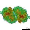 30547MC  7d1uC M: map data used to model this data C: citing same article ( |
|---|---|
| Similar structure data | |
| EM raw data | EMPIAR-10556 (Title: Cryo-EM Structure of PSII at 1.95 angstrom resolution Data size: 427.6 Data #1: Unaligned multi-frame micrographs of PSII recorded by CRYO ARM 300 [micrographs - multiframe]) |


-Links
 PDBj
PDBj


- Assembly
| Deposited unit | 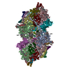
|
|---|---|
| 1 |
|
-Components
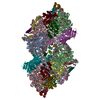
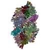
-Photosystem II ... , 17 types, 34 molecules AaBbCcDdHhIiJjKkLlMmOoTtUuYyXx...
| #1: Protein | Mass: 37029.234 Da / Num. of mol.: 2 / Source method: isolated from a natural source / Source: (natural) Thermosynechococcus vulcanus (bacteria) / References: UniProt: P51765, photosystem II#2: Protein | Mass: 56068.742 Da / Num. of mol.: 2 / Source method: isolated from a natural source / Source: (natural) Thermosynechococcus vulcanus (bacteria) / References: UniProt: D0VWR1#3: Protein | Mass: 49207.250 Da / Num. of mol.: 2 / Source method: isolated from a natural source / Source: (natural) Thermosynechococcus vulcanus (bacteria) / References: UniProt: D0VWR7#4: Protein | Mass: 38419.941 Da / Num. of mol.: 2 / Source method: isolated from a natural source / Source: (natural) Thermosynechococcus vulcanus (bacteria) / References: UniProt: D0VWR8, photosystem II#7: Protein | Mass: 7057.349 Da / Num. of mol.: 2 / Source method: isolated from a natural source / Source: (natural) Thermosynechococcus vulcanus (bacteria) / References: UniProt: P19052#8: Protein/peptide | Mass: 4195.983 Da / Num. of mol.: 2 / Source method: isolated from a natural source / Source: (natural) Thermosynechococcus vulcanus (bacteria) / References: UniProt: P12240#9: Protein/peptide | Mass: 3756.439 Da / Num. of mol.: 2 / Source method: isolated from a natural source / Source: (natural) Thermosynechococcus vulcanus (bacteria) / References: UniProt: Q7DGD4#10: Protein/peptide | Mass: 4101.911 Da / Num. of mol.: 2 / Source method: isolated from a natural source / Source: (natural) Thermosynechococcus vulcanus (bacteria) / References: UniProt: P19054#11: Protein/peptide | Mass: 4299.044 Da / Num. of mol.: 2 / Source method: isolated from a natural source / Source: (natural) Thermosynechococcus vulcanus (bacteria) / References: UniProt: P12241#12: Protein/peptide | Mass: 3835.527 Da / Num. of mol.: 2 / Source method: isolated from a natural source / Source: (natural) Thermosynechococcus vulcanus (bacteria) / References: UniProt: P12312#13: Protein | Mass: 26651.707 Da / Num. of mol.: 2 / Source method: isolated from a natural source / Source: (natural) Thermosynechococcus vulcanus (bacteria) / References: UniProt: D0VWR2#14: Protein/peptide | Mass: 3777.559 Da / Num. of mol.: 2 / Source method: isolated from a natural source / Source: (natural) Thermosynechococcus vulcanus (bacteria) / References: UniProt: P12313#15: Protein | Mass: 10966.317 Da / Num. of mol.: 2 / Source method: isolated from a natural source / Source: (natural) Thermosynechococcus vulcanus (bacteria) / References: UniProt: P56152#17: Protein/peptide | Mass: 3228.035 Da / Num. of mol.: 2 / Source method: isolated from a natural source / Source: (natural) Thermosynechococcus vulcanus (bacteria) / References: UniProt: D0VWR3#18: Protein/peptide | Mass: 4191.030 Da / Num. of mol.: 2 / Source method: isolated from a natural source / Source: (natural) Thermosynechococcus vulcanus (bacteria) / References: UniProt: D0VWR4#19: Protein | Mass: 6766.187 Da / Num. of mol.: 2 / Source method: isolated from a natural source / Source: (natural) Thermosynechococcus vulcanus (bacteria) / References: UniProt: D0VWR5#20: Protein/peptide | Mass: 3859.732 Da / Num. of mol.: 2 / Source method: isolated from a natural source / Source: (natural) Thermosynechococcus vulcanus (bacteria) / References: UniProt: P0DM37 |
|---|
-Cytochrome b559 subunit ... , 2 types, 4 molecules EeFf
| #5: Protein | Mass: 9321.515 Da / Num. of mol.: 2 / Source method: isolated from a natural source / Source: (natural) Thermosynechococcus vulcanus (bacteria) / References: UniProt: P12238#6: Protein/peptide | Mass: 3868.611 Da / Num. of mol.: 2 / Source method: isolated from a natural source / Source: (natural) Thermosynechococcus vulcanus (bacteria) / References: UniProt: P12239 |
|---|
-Protein , 1 types, 2 molecules Vv
| #16: Protein | Mass: 15148.255 Da / Num. of mol.: 2 / Source method: isolated from a natural source / Source: (natural) Thermosynechococcus vulcanus (bacteria) / References: UniProt: P0A387 |
|---|
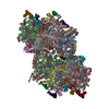
-Sugars , 2 types, 16 molecules 

| #31: Sugar | ChemComp-DGD /  Type: saccharide / Mass: 949.299 Da / Num. of mol.: 8 / Source method: obtained synthetically / Formula: C51H96O15 Type: saccharide / Mass: 949.299 Da / Num. of mol.: 8 / Source method: obtained synthetically / Formula: C51H96O15#32: Sugar | ChemComp-LMT /  Type: D-saccharide / Mass: 510.615 Da / Num. of mol.: 8 / Source method: obtained synthetically / Formula: C24H46O11 / Comment: detergent*YM Type: D-saccharide / Mass: 510.615 Da / Num. of mol.: 8 / Source method: obtained synthetically / Formula: C24H46O11 / Comment: detergent*YM |
|---|
-Non-polymers , 17 types, 2612 molecules 


| #21: Chemical |  Mass: 339.827 Da / Num. of mol.: 2 / Source method: obtained synthetically / Formula: CaMn4O5 Mass: 339.827 Da / Num. of mol.: 2 / Source method: obtained synthetically / Formula: CaMn4O5#22: Chemical |  Mass: 55.845 Da / Num. of mol.: 2 / Source method: obtained synthetically / Formula: Fe Mass: 55.845 Da / Num. of mol.: 2 / Source method: obtained synthetically / Formula: Fe#23: Chemical | ChemComp-CL /  Mass: 35.453 Da / Num. of mol.: 4 / Source method: obtained synthetically / Formula: Cl Mass: 35.453 Da / Num. of mol.: 4 / Source method: obtained synthetically / Formula: Cl#24: Chemical | ChemComp-CLA /  Mass: 893.489 Da / Num. of mol.: 70 / Source method: obtained synthetically / Formula: C55H72MgN4O5 Mass: 893.489 Da / Num. of mol.: 70 / Source method: obtained synthetically / Formula: C55H72MgN4O5#25: Chemical | ChemComp-PHO /  Mass: 871.200 Da / Num. of mol.: 4 / Source method: obtained synthetically / Formula: C55H74N4O5 Mass: 871.200 Da / Num. of mol.: 4 / Source method: obtained synthetically / Formula: C55H74N4O5#26: Chemical | ChemComp-BCR /  Mass: 536.873 Da / Num. of mol.: 20 / Source method: obtained synthetically / Formula: C40H56 Mass: 536.873 Da / Num. of mol.: 20 / Source method: obtained synthetically / Formula: C40H56#27: Chemical | ChemComp-SQD /  Mass: 795.116 Da / Num. of mol.: 8 / Source method: obtained synthetically / Formula: C41H78O12S Mass: 795.116 Da / Num. of mol.: 8 / Source method: obtained synthetically / Formula: C41H78O12S#28: Chemical | ChemComp-PL9 /  Mass: 749.201 Da / Num. of mol.: 4 / Source method: obtained synthetically / Formula: C53H80O2 Mass: 749.201 Da / Num. of mol.: 4 / Source method: obtained synthetically / Formula: C53H80O2#29: Chemical | ChemComp-UNL / Num. of mol.: 36 / Source method: obtained synthetically #30: Chemical | ChemComp-LMG /  Mass: 787.158 Da / Num. of mol.: 10 / Source method: obtained synthetically / Formula: C45H86O10 Mass: 787.158 Da / Num. of mol.: 10 / Source method: obtained synthetically / Formula: C45H86O10#33: Chemical |  Mass: 61.017 Da / Num. of mol.: 2 / Source method: obtained synthetically / Formula: CHO3 / Comment: pH buffer*YM Mass: 61.017 Da / Num. of mol.: 2 / Source method: obtained synthetically / Formula: CHO3 / Comment: pH buffer*YM#34: Chemical | ChemComp-LHG /  Mass: 722.970 Da / Num. of mol.: 10 / Source method: obtained synthetically / Formula: C38H75O10P / Comment: phospholipid*YM Mass: 722.970 Da / Num. of mol.: 10 / Source method: obtained synthetically / Formula: C38H75O10P / Comment: phospholipid*YM#35: Chemical |  Mass: 616.487 Da / Num. of mol.: 2 / Source method: obtained synthetically / Formula: C34H32FeN4O4 Mass: 616.487 Da / Num. of mol.: 2 / Source method: obtained synthetically / Formula: C34H32FeN4O4#36: Chemical |  Mass: 24.305 Da / Num. of mol.: 2 / Source method: isolated from a natural source / Formula: Mg Mass: 24.305 Da / Num. of mol.: 2 / Source method: isolated from a natural source / Formula: Mg#37: Chemical |  Mass: 618.503 Da / Num. of mol.: 2 / Source method: obtained synthetically / Formula: C34H34FeN4O4 Mass: 618.503 Da / Num. of mol.: 2 / Source method: obtained synthetically / Formula: C34H34FeN4O4#38: Chemical |  Mass: 552.872 Da / Num. of mol.: 2 / Source method: obtained synthetically / Formula: C40H56O Mass: 552.872 Da / Num. of mol.: 2 / Source method: obtained synthetically / Formula: C40H56O#39: Water | ChemComp-HOH / | Mass: 18.015 Da / Num. of mol.: 2432 / Source method: isolated from a natural source / Formula: H2O |
|---|
-Details
| Has ligand of interest | N |
|---|---|
| Has protein modification | Y |
-Experimental details
-Experiment
| Experiment | Method: ELECTRON MICROSCOPY |
|---|---|
| EM experiment | Aggregation state: PARTICLE / 3D reconstruction method: single particle reconstruction |
- Sample preparation
| Component | Name: PSII dimer / Type: COMPLEX / Entity ID: #1-#20 / Source: NATURAL | ||||||||||||
|---|---|---|---|---|---|---|---|---|---|---|---|---|---|
| Molecular weight | Value: 0.725 MDa / Experimental value: NO | ||||||||||||
| Source (natural) | Organism: Thermosynechococcus vulcanus (bacteria) | ||||||||||||
| Buffer solution | pH: 6 | ||||||||||||
| Buffer component |
| ||||||||||||
| Specimen | Conc.: 2 mg/ml / Embedding applied: NO / Shadowing applied: NO / Staining applied: NO / Vitrification applied: YES | ||||||||||||
| Specimen support | Grid material: COPPER / Grid mesh size: 200 divisions/in. / Grid type: Quantifoil R1.2/1.3 | ||||||||||||
| Vitrification | Instrument: FEI VITROBOT MARK IV / Cryogen name: ETHANE / Humidity: 100 % / Chamber temperature: 277 K |
- Electron microscopy imaging
| Microscopy | Model: JEOL CRYO ARM 300 |
|---|---|
| Electron gun | Electron source:  FIELD EMISSION GUN / Accelerating voltage: 300 kV / Illumination mode: FLOOD BEAM FIELD EMISSION GUN / Accelerating voltage: 300 kV / Illumination mode: FLOOD BEAM |
| Electron lens | Mode: BRIGHT FIELD |
| Image recording | Electron dose: 83 e/Å2 / Detector mode: COUNTING / Film or detector model: GATAN K2 SUMMIT (4k x 4k) |
- Processing
| Software | Name: PHENIX / Version: 1.14_3260: / Classification: refinement | ||||||||||||||||||||
|---|---|---|---|---|---|---|---|---|---|---|---|---|---|---|---|---|---|---|---|---|---|
| EM software |
| ||||||||||||||||||||
| CTF correction | Type: PHASE FLIPPING AND AMPLITUDE CORRECTION | ||||||||||||||||||||
| Symmetry | Point symmetry: C2 (2 fold cyclic) | ||||||||||||||||||||
| 3D reconstruction | Resolution: 1.95 Å / Resolution method: FSC 0.143 CUT-OFF / Num. of particles: 174099 / Algorithm: FOURIER SPACE / Symmetry type: POINT | ||||||||||||||||||||
| Atomic model building | Protocol: FLEXIBLE FIT / Space: REAL | ||||||||||||||||||||
| Atomic model building | PDB-ID: 3WU2 Accession code: 3WU2 / Source name: PDB / Type: experimental model |