 Movie
Movie Controller


+ Open data

- Basic information
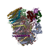
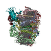
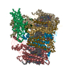
| Entry | Database: PDB / ID: 7eda | ||||||||||||||||||||||||||||||||||||||||||
|---|---|---|---|---|---|---|---|---|---|---|---|---|---|---|---|---|---|---|---|---|---|---|---|---|---|---|---|---|---|---|---|---|---|---|---|---|---|---|---|---|---|---|---|
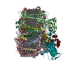
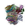
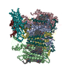
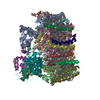
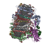
| Title | Structure of monomeric photosystem II | ||||||||||||||||||||||||||||||||||||||||||
 Components Components |
| ||||||||||||||||||||||||||||||||||||||||||
 Keywords Keywords | PHOTOSYNTHESIS / Photosystem / membrane protein | ||||||||||||||||||||||||||||||||||||||||||
| Function / homology |  Function and homology information Function and homology informationphotosystem II oxygen evolving complex / photosystem II assembly / oxygen evolving activity / photosystem II stabilization / photosystem II reaction center / photosystem II / photosynthetic electron transport chain / oxidoreductase activity, acting on diphenols and related substances as donors, oxygen as acceptor / response to herbicide / photosystem II ...photosystem II oxygen evolving complex / photosystem II assembly / oxygen evolving activity / photosystem II stabilization / photosystem II reaction center / photosystem II / photosynthetic electron transport chain / oxidoreductase activity, acting on diphenols and related substances as donors, oxygen as acceptor / response to herbicide / photosystem II / extrinsic component of membrane / plasma membrane-derived thylakoid membrane / photosynthetic electron transport in photosystem II / chlorophyll binding / photosynthesis, light reaction / phosphate ion binding / photosynthesis / respiratory electron transport chain / manganese ion binding / electron transfer activity / protein stabilization / iron ion binding / heme binding / metal ion binding Similarity search - Function | ||||||||||||||||||||||||||||||||||||||||||
| Biological species |  Thermosynechococcus vulcanus (bacteria) Thermosynechococcus vulcanus (bacteria) | ||||||||||||||||||||||||||||||||||||||||||
| Method | ELECTRON MICROSCOPY / single particle reconstruction / cryo EM / Resolution: 2.78 Å | ||||||||||||||||||||||||||||||||||||||||||
 Authors Authors | Yu, H. / Hamaguchi, T. / Nakajima, Y. / Kato, K. / kawakami, K. / Akita, F. / Yonekura, K. / Shen, J.R. | ||||||||||||||||||||||||||||||||||||||||||
| Funding support |  Japan, 6items Japan, 6items
| ||||||||||||||||||||||||||||||||||||||||||
 Citation Citation | Journal: Biochim Biophys Acta Bioenerg / Year: 2021 Title: Cryo-EM structure of monomeric photosystem II at 2.78 Å resolution reveals factors important for the formation of dimer. Authors: Huaxin Yu / Tasuku Hamaguchi / Yoshiki Nakajima / Koji Kato / Keisuke Kawakami / Fusamichi Akita / Koji Yonekura / Jian-Ren Shen / Abstract: Photosystem II (PSII) functions mainly as a dimer to catalyze the light energy conversion and water oxidation reactions. However, monomeric PSII also exists and functions in vivo in some cases. The ...Photosystem II (PSII) functions mainly as a dimer to catalyze the light energy conversion and water oxidation reactions. However, monomeric PSII also exists and functions in vivo in some cases. The crystal structure of monomeric PSII has been solved at 3.6 Å resolution, but it is still not clear which factors contribute to the formation of the dimer. Here, we solved the structure of PSII monomer at a resolution of 2.78 Å using cryo-electron microscopy (cryo-EM). From our cryo-EM density map, we observed apparent differences in pigments and lipids in the monomer-monomer interface between the PSII monomer and dimer. One β-carotene and two sulfoquinovosyl diacylglycerol (SQDG) molecules are found in the monomer-monomer interface of the dimer structure but not in the present monomer structure, although some SQDG and other lipid molecules are found in the analogous region of the low-resolution crystal structure of the monomer, or cryo-EM structure of an apo-PSII monomer lacking the extrinsic proteins from Synechocystis sp. PCC 6803. In the current monomer structure, a large part of the PsbO subunit was also found to be disordered. These results indicate the importance of the β-carotene, SQDG and PsbO in formation of the PSII dimer. | ||||||||||||||||||||||||||||||||||||||||||
| History |
|
- Structure visualization
| Movie |
Movie viewer |
|---|---|
| Structure viewer | Molecule: MolmilJmol/JSmol |
- Downloads & links
-Download
| PDBx/mmCIF format | 7eda.cif.gz | 545.3 KB | Display | PDBx/mmCIF format |
|---|---|---|---|---|
| PDB format | pdb7eda.ent.gz | 437 KB | Display | PDB format |
| PDBx/mmJSON format | 7eda.json.gz | Tree view | PDBx/mmJSON format | |
| Others |  Other downloads Other downloads |
-Validation report
| Arichive directory | https://data.pdbj.org/pub/pdb/validation_reports/ed/7edaftp://data.pdbj.org/pub/pdb/validation_reports/ed/7eda | HTTPS FTP |
|---|
-Related structure data
| Related structure data | 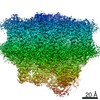 31062MC M: map data used to model this data C: citing same article ( |
|---|---|
| Similar structure data |


-Links
 PDBj
PDBj


- Assembly
| Deposited unit | 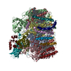
|
|---|---|
| 1 |
|
-Components
-Photosystem II ... , 17 types, 17 molecules ABCDHIJKLMOTUYXZR
| #1: Protein | Mass: 37029.234 Da / Num. of mol.: 1 / Source method: isolated from a natural source / Source: (natural) Thermosynechococcus vulcanus (bacteria) / References: UniProt: P51765, photosystem II |
|---|---|
| #2: Protein | Mass: 55939.562 Da / Num. of mol.: 1 / Source method: isolated from a natural source / Source: (natural) Thermosynechococcus vulcanus (bacteria) / References: UniProt: D0VWR1 |
| #3: Protein | Mass: 49207.250 Da / Num. of mol.: 1 / Source method: isolated from a natural source / Source: (natural) Thermosynechococcus vulcanus (bacteria) / References: UniProt: D0VWR7 |
| #4: Protein | Mass: 38290.828 Da / Num. of mol.: 1 / Source method: isolated from a natural source / Source: (natural) Thermosynechococcus vulcanus (bacteria) / References: UniProt: D0VWR8, photosystem II |
| #7: Protein | Mass: 6986.271 Da / Num. of mol.: 1 / Source method: isolated from a natural source / Source: (natural) Thermosynechococcus vulcanus (bacteria) / References: UniProt: P19052 |
| #8: Protein/peptide | Mass: 4438.255 Da / Num. of mol.: 1 / Source method: isolated from a natural source / Source: (natural) Thermosynechococcus vulcanus (bacteria) / References: UniProt: P12240 |
| #9: Protein/peptide | Mass: 4105.908 Da / Num. of mol.: 1 / Source method: isolated from a natural source / Source: (natural) Thermosynechococcus vulcanus (bacteria) / References: UniProt: Q7DGD4 |
| #10: Protein/peptide | Mass: 4101.911 Da / Num. of mol.: 1 / Source method: isolated from a natural source / Source: (natural) Thermosynechococcus vulcanus (bacteria) / References: UniProt: P19054 |
| #11: Protein/peptide | Mass: 4299.044 Da / Num. of mol.: 1 / Source method: isolated from a natural source / Source: (natural) Thermosynechococcus vulcanus (bacteria) / References: UniProt: P12241 |
| #12: Protein/peptide | Mass: 3289.899 Da / Num. of mol.: 1 / Source method: isolated from a natural source / Source: (natural) Thermosynechococcus vulcanus (bacteria) / References: UniProt: P12312 |
| #13: Protein | Mass: 26651.707 Da / Num. of mol.: 1 / Source method: isolated from a natural source / Source: (natural) Thermosynechococcus vulcanus (bacteria) / References: UniProt: D0VWR2 |
| #14: Protein/peptide | Mass: 3648.379 Da / Num. of mol.: 1 / Source method: isolated from a natural source / Source: (natural) Thermosynechococcus vulcanus (bacteria) / References: UniProt: P12313 |
| #15: Protein | Mass: 11655.986 Da / Num. of mol.: 1 / Source method: isolated from a natural source / Source: (natural) Thermosynechococcus vulcanus (bacteria) / References: UniProt: P56152 |
| #17: Protein/peptide | Mass: 3228.035 Da / Num. of mol.: 1 / Source method: isolated from a natural source / Source: (natural) Thermosynechococcus vulcanus (bacteria) / References: UniProt: D0VWR3 |
| #18: Protein/peptide | Mass: 4191.030 Da / Num. of mol.: 1 / Source method: isolated from a natural source / Source: (natural) Thermosynechococcus vulcanus (bacteria) / References: UniProt: D0VWR4 |
| #19: Protein | Mass: 6766.187 Da / Num. of mol.: 1 / Source method: isolated from a natural source / Source: (natural) Thermosynechococcus vulcanus (bacteria) / References: UniProt: D0VWR5 |
| #20: Protein/peptide | Mass: 3859.732 Da / Num. of mol.: 1 / Source method: isolated from a natural source / Source: (natural) Thermosynechococcus vulcanus (bacteria) / References: UniProt: P0DM37 |
-Cytochrome b559 subunit ... , 2 types, 2 molecules EF
| #5: Protein | Mass: 9580.840 Da / Num. of mol.: 1 / Source method: isolated from a natural source / Source: (natural) Thermosynechococcus vulcanus (bacteria) / References: UniProt: P12238 |
|---|---|
| #6: Protein/peptide | Mass: 5067.900 Da / Num. of mol.: 1 / Source method: isolated from a natural source / Source: (natural) Thermosynechococcus vulcanus (bacteria) / References: UniProt: P12239 |
-Protein / Sugars , 2 types, 5 molecules V
| #16: Protein | Mass: 18046.943 Da / Num. of mol.: 1 / Source method: isolated from a natural source / Source: (natural) Thermosynechococcus vulcanus (bacteria) / References: UniProt: P0A387 |
|---|---|
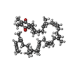
| #30: Sugar | ChemComp-DGD /  Type: saccharide / Mass: 949.299 Da / Num. of mol.: 4 / Source method: obtained synthetically / Formula: C51H96O15 / Feature type: SUBJECT OF INVESTIGATION Type: saccharide / Mass: 949.299 Da / Num. of mol.: 4 / Source method: obtained synthetically / Formula: C51H96O15 / Feature type: SUBJECT OF INVESTIGATION |
-Non-polymers , 15 types, 79 molecules 


| #21: Chemical | ChemComp-OEX /  Mass: 339.827 Da / Num. of mol.: 1 / Source method: obtained synthetically / Formula: CaMn4O5 Mass: 339.827 Da / Num. of mol.: 1 / Source method: obtained synthetically / Formula: CaMn4O5 | ||||||||||||||||||||||||
|---|---|---|---|---|---|---|---|---|---|---|---|---|---|---|---|---|---|---|---|---|---|---|---|---|---|
| #22: Chemical | ChemComp-FE2 /  Mass: 55.845 Da / Num. of mol.: 1 / Source method: obtained synthetically / Formula: Fe Mass: 55.845 Da / Num. of mol.: 1 / Source method: obtained synthetically / Formula: Fe | ||||||||||||||||||||||||
| #23: Chemical | ChemComp-CLA /  Mass: 893.489 Da / Num. of mol.: 35 / Source method: obtained synthetically / Formula: C55H72MgN4O5 Mass: 893.489 Da / Num. of mol.: 35 / Source method: obtained synthetically / Formula: C55H72MgN4O5#24: Chemical |  Mass: 871.200 Da / Num. of mol.: 2 / Source method: obtained synthetically / Formula: C55H74N4O5 Mass: 871.200 Da / Num. of mol.: 2 / Source method: obtained synthetically / Formula: C55H74N4O5#25: Chemical | ChemComp-BCR /  Mass: 536.873 Da / Num. of mol.: 8 / Source method: obtained synthetically / Formula: C40H56 / Feature type: SUBJECT OF INVESTIGATION Mass: 536.873 Da / Num. of mol.: 8 / Source method: obtained synthetically / Formula: C40H56 / Feature type: SUBJECT OF INVESTIGATION#26: Chemical |  Mass: 795.116 Da / Num. of mol.: 2 / Source method: obtained synthetically / Formula: C41H78O12S / Feature type: SUBJECT OF INVESTIGATION Mass: 795.116 Da / Num. of mol.: 2 / Source method: obtained synthetically / Formula: C41H78O12S / Feature type: SUBJECT OF INVESTIGATION#27: Chemical |  Mass: 787.158 Da / Num. of mol.: 3 / Source method: obtained synthetically / Formula: C45H86O10 / Feature type: SUBJECT OF INVESTIGATION Mass: 787.158 Da / Num. of mol.: 3 / Source method: obtained synthetically / Formula: C45H86O10 / Feature type: SUBJECT OF INVESTIGATION#28: Chemical |  Mass: 749.201 Da / Num. of mol.: 2 / Source method: obtained synthetically / Formula: C53H80O2 Mass: 749.201 Da / Num. of mol.: 2 / Source method: obtained synthetically / Formula: C53H80O2#29: Chemical | ChemComp-UNL / | Mass: 949.299 Da / Num. of mol.: 1 / Source method: obtained synthetically / Feature type: SUBJECT OF INVESTIGATION #31: Chemical | ChemComp-BCT / |  Mass: 61.017 Da / Num. of mol.: 1 / Source method: obtained synthetically / Formula: CHO3 / Comment: pH buffer*YM Mass: 61.017 Da / Num. of mol.: 1 / Source method: obtained synthetically / Formula: CHO3 / Comment: pH buffer*YM#32: Chemical | ChemComp-LHG /  Mass: 722.970 Da / Num. of mol.: 4 / Source method: obtained synthetically / Formula: C38H75O10P / Feature type: SUBJECT OF INVESTIGATION / Comment: phospholipid*YM Mass: 722.970 Da / Num. of mol.: 4 / Source method: obtained synthetically / Formula: C38H75O10P / Feature type: SUBJECT OF INVESTIGATION / Comment: phospholipid*YM#33: Chemical | ChemComp-HEM / |  Mass: 616.487 Da / Num. of mol.: 1 / Source method: isolated from a natural source / Formula: C34H32FeN4O4 Mass: 616.487 Da / Num. of mol.: 1 / Source method: isolated from a natural source / Formula: C34H32FeN4O4#34: Chemical | ChemComp-RRX / ( |  Mass: 552.872 Da / Num. of mol.: 1 / Source method: obtained synthetically / Formula: C40H56O / Feature type: SUBJECT OF INVESTIGATION Mass: 552.872 Da / Num. of mol.: 1 / Source method: obtained synthetically / Formula: C40H56O / Feature type: SUBJECT OF INVESTIGATION#35: Chemical | ChemComp-HEC / |  Mass: 618.503 Da / Num. of mol.: 1 / Source method: isolated from a natural source / Formula: C34H34FeN4O4 Mass: 618.503 Da / Num. of mol.: 1 / Source method: isolated from a natural source / Formula: C34H34FeN4O4#36: Water | ChemComp-HOH / | Mass: 18.015 Da / Num. of mol.: 16 / Source method: isolated from a natural source / Formula: H2O |
-Details
| Has ligand of interest | Y |
|---|---|
| Has protein modification | Y |
-Experimental details
-Experiment
| Experiment | Method: ELECTRON MICROSCOPY |
|---|---|
| EM experiment | Aggregation state: PARTICLE / 3D reconstruction method: single particle reconstruction |
- Sample preparation
| Component | Name: Photosystem II / Type: COMPLEX / Entity ID: #1-#6, #8-#20 / Source: NATURAL |
|---|---|
| Source (natural) | Organism: Thermosynechococcus vulcanus (bacteria) |
| Buffer solution | pH: 6 |
| Specimen | Embedding applied: NO / Shadowing applied: NO / Staining applied: NO / Vitrification applied: YES |
| Vitrification | Cryogen name: ETHANE |
- Electron microscopy imaging
| Microscopy | Model: JEOL CRYO ARM 300 |
|---|---|
| Electron gun | Electron source:  FIELD EMISSION GUN / Accelerating voltage: 300 kV / Illumination mode: FLOOD BEAM FIELD EMISSION GUN / Accelerating voltage: 300 kV / Illumination mode: FLOOD BEAM |
| Electron lens | Mode: BRIGHT FIELD |
| Image recording | Electron dose: 70 e/Å2 / Film or detector model: GATAN K3 (6k x 4k) |
- Processing
| Software | Name: PHENIX / Version: 1.14_3260: / Classification: refinement | ||||||||||||||||||||||||
|---|---|---|---|---|---|---|---|---|---|---|---|---|---|---|---|---|---|---|---|---|---|---|---|---|---|
| EM software | Name: PHENIX / Category: model refinement | ||||||||||||||||||||||||
| CTF correction | Type: NONE | ||||||||||||||||||||||||
| 3D reconstruction | Resolution: 2.78 Å / Resolution method: FSC 0.143 CUT-OFF / Num. of particles: 173875 / Symmetry type: POINT | ||||||||||||||||||||||||
| Refine LS restraints |
|
