 Movie
Movie Controller

[English] 日本語
 Yorodumi
- PDB-7vea: Pentacylindrical allophycocyanin core from Thermosynechococcus vu... -
+ Open data

- Basic information
| Entry | Database: PDB / ID: 7vea | ||||||||||||||||||||||||||||||
|---|---|---|---|---|---|---|---|---|---|---|---|---|---|---|---|---|---|---|---|---|---|---|---|---|---|---|---|---|---|---|---|
| Title | Pentacylindrical allophycocyanin core from Thermosynechococcus vulcanus | ||||||||||||||||||||||||||||||
 Components Components |
| ||||||||||||||||||||||||||||||
 Keywords Keywords | PHOTOSYNTHESIS / Light-harvesting complex / Phycobilisome / Energy transfer | ||||||||||||||||||||||||||||||
| Function / homology |  Function and homology information Function and homology informationphycobilisome / plasma membrane-derived thylakoid membrane / photosynthesis / lyase activity Similarity search - Function | ||||||||||||||||||||||||||||||
| Biological species |   Thermosynechococcus vestitus BP-1 (bacteria) Thermosynechococcus vestitus BP-1 (bacteria) | ||||||||||||||||||||||||||||||
| Method | ELECTRON MICROSCOPY / single particle reconstruction / cryo EM / Resolution: 3.7 Å | ||||||||||||||||||||||||||||||
 Authors Authors | Kawakami, K. / Hamaguchi, T. / Hirose, Y. / Kosumi, D. / Miyata, M. / Kamiya, N. / Yonekura, K. | ||||||||||||||||||||||||||||||
| Funding support |  Japan, 3items Japan, 3items
| ||||||||||||||||||||||||||||||
 Citation Citation | Journal: Nat Commun / Year: 2022 Title: Core and rod structures of a thermophilic cyanobacterial light-harvesting phycobilisome. Authors: Keisuke Kawakami / Tasuku Hamaguchi / Yuu Hirose / Daisuke Kosumi / Makoto Miyata / Nobuo Kamiya / Koji Yonekura / Abstract: Cyanobacteria, glaucophytes, and rhodophytes utilize giant, light-harvesting phycobilisomes (PBSs) for capturing solar energy and conveying it to photosynthetic reaction centers. PBSs are ...Cyanobacteria, glaucophytes, and rhodophytes utilize giant, light-harvesting phycobilisomes (PBSs) for capturing solar energy and conveying it to photosynthetic reaction centers. PBSs are compositionally and structurally diverse, and exceedingly complex, all of which pose a challenge for a comprehensive understanding of their function. To date, three detailed architectures of PBSs by cryo-electron microscopy (cryo-EM) have been described: a hemiellipsoidal type, a block-type from rhodophytes, and a cyanobacterial hemidiscoidal-type. Here, we report cryo-EM structures of a pentacylindrical allophycocyanin core and phycocyanin-containing rod of a thermophilic cyanobacterial hemidiscoidal PBS. The structures define the spatial arrangement of protein subunits and chromophores, crucial for deciphering the energy transfer mechanism. They reveal how the pentacylindrical core is formed, identify key interactions between linker proteins and the bilin chromophores, and indicate pathways for unidirectional energy transfer. | ||||||||||||||||||||||||||||||
| History |
|
- Structure visualization
| Structure viewer | Molecule: MolmilJmol/JSmol |
|---|
- Downloads & links
-Download
| PDBx/mmCIF format | 7vea.cif.gz | 3.2 MB | Display | PDBx/mmCIF format |
|---|---|---|---|---|
| PDB format | pdb7vea.ent.gz | Display | PDB format | |
| PDBx/mmJSON format | 7vea.json.gz | Tree view | PDBx/mmJSON format | |
| Others |  Other downloads Other downloads |
-Validation report
| Arichive directory | https://data.pdbj.org/pub/pdb/validation_reports/ve/7veaftp://data.pdbj.org/pub/pdb/validation_reports/ve/7vea | HTTPS FTP |
|---|
-Related structure data
| Related structure data | 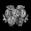 31944MC  7vebC M: map data used to model this data C: citing same article ( |
|---|---|
| Similar structure data |

-Links
 PDBj
PDBj- Assembly
| Deposited unit | 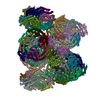
|
|---|---|
| 1 |
|
-Components
-Allophycocyanin ... , 2 types, 80 molecules aAaCaEaGaIaKaOaQbMbObQbSbUbWcAcCcEcGcIcKdAdCdEdGdIdKdOdQeMeO...
| #1: Protein | Mass: 17555.926 Da / Num. of mol.: 40 / Source method: isolated from a natural source Source: (natural) Thermosynechococcus vestitus BP-1 (bacteria)Strain: BP-1 / References: UniProt: P50030 #2: Protein | Mass: 17388.877 Da / Num. of mol.: 40 / Source method: isolated from a natural source Source: (natural) Thermosynechococcus vestitus BP-1 (bacteria)Strain: BP-1 / References: UniProt: P50031 |
|---|
-Phycobilisome ... , 2 types, 8 molecules aRdRaSbYcMdSeYfM
| #4: Protein | Mass: 18757.221 Da / Num. of mol.: 2 / Source method: isolated from a natural source Source: (natural) Thermosynechococcus vestitus BP-1 (bacteria)Strain: BP-1 / References: UniProt: Q8DHC5 #5: Protein | Mass: 7876.255 Da / Num. of mol.: 6 / Source method: isolated from a natural source Source: (natural) Thermosynechococcus vestitus BP-1 (bacteria)Strain: BP-1 / References: UniProt: P50036 |
|---|
-Protein / Non-polymers , 2 types, 86 molecules aMdM
| #3: Protein | Mass: 127772.922 Da / Num. of mol.: 2 / Source method: isolated from a natural source Source: (natural) Thermosynechococcus vestitus BP-1 (bacteria)Strain: BP-1 / References: UniProt: Q8DGF2 #6: Chemical | ChemComp-CYC /  Mass: 588.694 Da / Num. of mol.: 84 / Source method: obtained synthetically / Formula: C33H40N4O6 / Feature type: SUBJECT OF INVESTIGATION Mass: 588.694 Da / Num. of mol.: 84 / Source method: obtained synthetically / Formula: C33H40N4O6 / Feature type: SUBJECT OF INVESTIGATION |
|---|
-Details
| Has ligand of interest | Y |
|---|---|
| Has protein modification | Y |
-Experimental details
-Experiment
| Experiment | Method: ELECTRON MICROSCOPY |
|---|---|
| EM experiment | Aggregation state: PARTICLE / 3D reconstruction method: single particle reconstruction |
- Sample preparation
| Component | Name: Phycobilisome core complex from Thermosynechococcus vulcanus Type: COMPLEX / Entity ID: #1-#5 / Source: NATURAL |
|---|---|
| Source (natural) | Organism: Thermosynechococcus vulcanus NIES-2134 (bacteria) |
| Buffer solution | pH: 6.5 |
| Specimen | Embedding applied: NO / Shadowing applied: NO / Staining applied: NO / Vitrification applied: YES |
| Vitrification | Cryogen name: NITROGEN |
- Electron microscopy imaging
| Microscopy | Model: JEOL CRYO ARM 300 |
|---|---|
| Electron gun | Electron source:  FIELD EMISSION GUN / Accelerating voltage: 300 kV / Illumination mode: FLOOD BEAM FIELD EMISSION GUN / Accelerating voltage: 300 kV / Illumination mode: FLOOD BEAM |
| Electron lens | Mode: BRIGHT FIELD / Nominal defocus max: 1500 nm / Nominal defocus min: 500 nm / Cs: 2.7 mm / C2 aperture diameter: 150 µm |
| Image recording | Electron dose: 84.1 e/Å2 / Film or detector model: GATAN K2 SUMMIT (4k x 4k) |
- Processing
| Software |
| ||||||||||||||||||||||||
|---|---|---|---|---|---|---|---|---|---|---|---|---|---|---|---|---|---|---|---|---|---|---|---|---|---|
| EM software |
| ||||||||||||||||||||||||
| CTF correction | Type: PHASE FLIPPING AND AMPLITUDE CORRECTION | ||||||||||||||||||||||||
| 3D reconstruction | Resolution: 3.7 Å / Resolution method: FSC 0.143 CUT-OFF / Num. of particles: 25532 / Symmetry type: POINT | ||||||||||||||||||||||||
| Refinement | Cross valid method: NONE Stereochemistry target values: GeoStd + Monomer Library + CDL v1.2 | ||||||||||||||||||||||||
| Displacement parameters | Biso mean: 144.5 Å2 | ||||||||||||||||||||||||
| Refine LS restraints |
|
