 Movie
Movie Controller
 Structure viewers
 About Yorodumi Papers


+Search query
-Structure paper
| Title | Protein and Organic-Molecular Crystallography With 300kV Electrons on a Direct Electron Detector. |
|---|---|
| Journal, issue, pages | Front Mol Biosci, Vol. 7, Page 612226, Year 2020 |
| Publish date | Jan 6, 2021 |
 Authors Authors | Kiyofumi Takaba / Saori Maki-Yonekura / Satoru Inoue / Tatsuo Hasegawa / Koji Yonekura /  |
| PubMed Abstract | Electron 3D crystallography can reveal the atomic structure from undersized crystals of various samples owing to the strong scattering power of electrons. Here, a direct electron detector DE64 was ...Electron 3D crystallography can reveal the atomic structure from undersized crystals of various samples owing to the strong scattering power of electrons. Here, a direct electron detector DE64 was tested for small and thin crystals of protein and an organic molecule using a JEOL CRYO ARM 300 electron microscope. The microscope is equipped with a cold-field emission gun operated at an accelerating voltage of 300 kV, quad condenser lenses for parallel illumination, an in-column energy filter, and a stable rotational goniometer stage. Rotational diffraction data were collected in an unsupervised manner from crystals of a heme-binding enzyme catalase and a representative organic semiconductor material Ph-BTBT-C10. The structures were determined by molecular replacement for catalase and by the direct method for Ph-BTBT-C10. The analyses demonstrate that the system works well for electron 3D crystallography of these molecules with less damaging, a smaller point spread, and less noise than using the conventional scintillator-coupled camera. |
 External links External links | Front Mol Biosci / PubMed:33469549 / PubMed Central |
| Methods | EM (electron crystallography) |
| Resolution | 3.2 Å |
| Structure data | 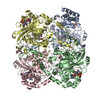 PDB-7di8: |
| Chemicals |  ChemComp-HEM:  ChemComp-NDP: |
| Source |
|
 Keywords Keywords | OXIDOREDUCTASE / electron 3d crystallography / direct detector / cryo arm / parallem |

