Journal: J Mol Biol / Year: 2004 Title: Structure of the rotor of the bacterial flagellar motor revealed by electron cryomicroscopy and single-particle image analysis. Authors: Hirofumi Suzuki / Koji Yonekura / Keiichi Namba / Abstract: The FliF ring is the base for self-assembly of the bacterial flagellum and the FliF/FliG ring complex is the core of the rotor of the flagellar motor. We report the structures of these two ring ...The FliF ring is the base for self-assembly of the bacterial flagellum and the FliF/FliG ring complex is the core of the rotor of the flagellar motor. We report the structures of these two ring complexes obtained by electron cryomicroscopy and single-particle image analysis at 22A and 25A resolution, respectively. Direct comparison of these structures with the flagellar basal body made by superimposing the density maps on the central section reveals many interesting features, such as how the mechanically stable connection between the ring and the rod is formed, how directly FliF domains are involved in the near axial density of the basal body forming the proximal end of the central channel for a potential gating mechanism, some indication of flexibility in the connection of FliF and FliG, and structural and functional similarities to the head-to-tail connectors of bacteriophages.
History
Deposition
Mar 16, 2017
-
Header (metadata) release
Apr 12, 2017
-
Map release
Apr 12, 2017
-
Update
Apr 12, 2017
-
Current status
Apr 12, 2017
Processing site: PDBj / Status: Released
-
Structure visualization
Movie
Surface view with section colored by density value
In the structure databanks used in Yorodumi, some data are registered as the other names, "COVID-19 virus" and "2019-nCoV". Here are the details of the virus and the list of structure data.
Jan 31, 2019. EMDB accession codes are about to change! (news from PDBe EMDB page)
EMDB accession codes are about to change! (news from PDBe EMDB page)
The allocation of 4 digits for EMDB accession codes will soon come to an end. Whilst these codes will remain in use, new EMDB accession codes will include an additional digit and will expand incrementally as the available range of codes is exhausted. The current 4-digit format prefixed with “EMD-” (i.e. EMD-XXXX) will advance to a 5-digit format (i.e. EMD-XXXXX), and so on. It is currently estimated that the 4-digit codes will be depleted around Spring 2019, at which point the 5-digit format will come into force.
The EM Navigator/Yorodumi systems omit the EMD- prefix.
Related info.:Q: What is EMD? / ID/Accession-code notation in Yorodumi/EM Navigator
Yorodumi is a browser for structure data from EMDB, PDB, SASBDB, etc.
This page is also the successor to EM Navigator detail page, and also detail information page/front-end page for Omokage search.
The word "yorodu" (or yorozu) is an old Japanese word meaning "ten thousand". "mi" (miru) is to see.
Related info.:EMDB / PDB / SASBDB / Comparison of 3 databanks / Yorodumi Search / Aug 31, 2016. New EM Navigator & Yorodumi / Yorodumi Papers / Jmol/JSmol / Function and homology information / Changes in new EM Navigator and Yorodumi
 Movie
Movie Controller


 Open data
 Basic information
 Map data
 Sample
 Function and homology information
 Salmonella enterica subsp. enterica serovar Typhimurium (bacteria)
 Authors
 Citation
 Structure visualization
 Downloads & links
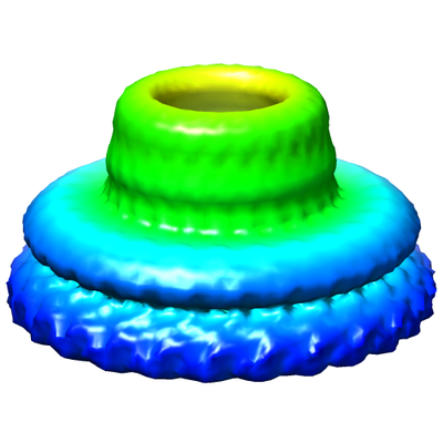 emd_6716.png
 http://ftp.pdbj.org/pub/emdb/structures/EMD-6716


 Z (Sec.)
 Y (Row.)
 X (Col.)


 Sample components
 Processing
 Electron microscopy
 FIELD EMISSION GUN