 Movie
Movie Controller


+ Open data

- Basic information
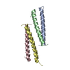
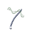
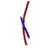
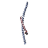
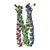
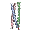
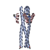
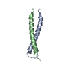
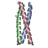
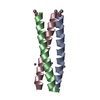
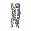
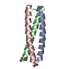
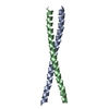
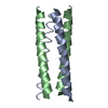
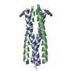
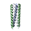
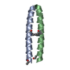
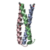
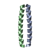
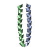
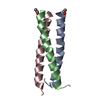
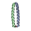
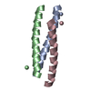
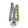
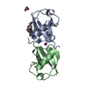
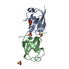
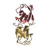
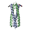
| Entry | Database: PDB / ID: 2bni | ||||||
|---|---|---|---|---|---|---|---|
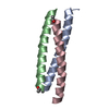
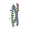
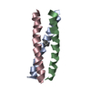
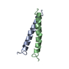
| Title | pLI mutant E20C L16G Y17H, antiparallel | ||||||
 Components Components | GENERAL CONTROL PROTEIN GCN4 | ||||||
 Keywords Keywords | FOUR HELIX BUNDLE / ANTIPARALLEL FOUR HELIX BUNDLE ACYL TRANSFERASE | ||||||
| Function / homology |  Function and homology information Function and homology informationFCERI mediated MAPK activation / protein localization to nuclear periphery / Activation of the AP-1 family of transcription factors / negative regulation of ribosomal protein gene transcription by RNA polymerase II / positive regulation of cellular response to amino acid starvation / response to amino acid starvation / mediator complex binding / Oxidative Stress Induced Senescence / amino acid biosynthetic process / TFIID-class transcription factor complex binding ...FCERI mediated MAPK activation / protein localization to nuclear periphery / Activation of the AP-1 family of transcription factors / negative regulation of ribosomal protein gene transcription by RNA polymerase II / positive regulation of cellular response to amino acid starvation / response to amino acid starvation / mediator complex binding / Oxidative Stress Induced Senescence / amino acid biosynthetic process / TFIID-class transcription factor complex binding / positive regulation of RNA polymerase II transcription preinitiation complex assembly / positive regulation of transcription initiation by RNA polymerase II / cellular response to nutrient levels / cellular response to amino acid starvation / RNA polymerase II transcription regulator complex / DNA-binding transcription activator activity, RNA polymerase II-specific / transcription regulator complex / sequence-specific DNA binding / RNA polymerase II-specific DNA-binding transcription factor binding / DNA-binding transcription factor activity, RNA polymerase II-specific / intracellular signal transduction / RNA polymerase II cis-regulatory region sequence-specific DNA binding / DNA-binding transcription factor activity / chromatin binding / negative regulation of transcription by RNA polymerase II / positive regulation of transcription by RNA polymerase II / identical protein binding / nucleus Similarity search - Function | ||||||
| Biological species |  | ||||||
| Method |  X-RAY DIFFRACTION / SYNCHROTRON / MOLECULAR REPLACEMENT / Resolution: 1.5 Å X-RAY DIFFRACTION / SYNCHROTRON / MOLECULAR REPLACEMENT / Resolution: 1.5 Å | ||||||
 Authors Authors | Yadav, M.K. / Leman, L.J. / Stout, C.D. / Ghadiri, M.R. | ||||||
 Citation Citation | Journal: Biochemistry / Year: 2005 Title: Structure-Based Engineering of Internal Cavities in Coiled-Coil Peptides Authors: Yadav, M.K. / Redman, J.E. / Leman, L.J. / Alvarez-Gutierrez, J.M. / Zhang, Y. / Stout, C.D. / Ghadiri, M.R. | ||||||
| History |
|
- Structure visualization
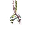
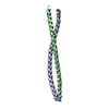
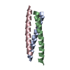
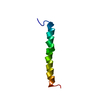
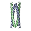
| Structure viewer | Molecule: MolmilJmol/JSmol |
|---|
- Downloads & links
-Download
| PDBx/mmCIF format | 2bni.cif.gz | 38.8 KB | Display | PDBx/mmCIF format |
|---|---|---|---|---|
| PDB format | pdb2bni.ent.gz | 28.3 KB | Display | PDB format |
| PDBx/mmJSON format | 2bni.json.gz | Tree view | PDBx/mmJSON format | |
| Others |  Other downloads Other downloads |
-Validation report
| Arichive directory | https://data.pdbj.org/pub/pdb/validation_reports/bn/2bniftp://data.pdbj.org/pub/pdb/validation_reports/bn/2bni | HTTPS FTP |
|---|
-Related structure data
| Related structure data | 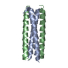 1untC 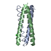 1unuC 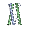 1unvC 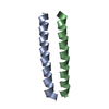 1unwC 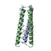 1unxC 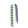 1unyC 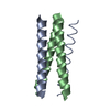 1unzC 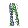 1uo0C 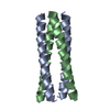 1uo1C 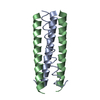 1uo2C 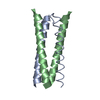 1uo3C  1uo4C 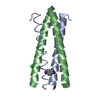 1uo5C 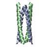 1w5gC 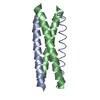 1w5iC C: citing same article ( |
|---|---|
| Similar structure data |


-Links
 PDBj
PDBj

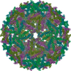
- Assembly
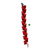
| Deposited unit | 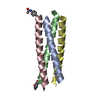
| ||||||||
|---|---|---|---|---|---|---|---|---|---|
| 1 |
| ||||||||
| Unit cell |
|
-Components
| #1: Protein/peptide | Mass: 4100.854 Da / Num. of mol.: 4 / Mutation: YES / Source method: obtained synthetically / Source: (synth.) #2: Water | ChemComp-HOH / |  Mass: 18.015 Da / Num. of mol.: 55 / Source method: isolated from a natural source / Formula: H2O Mass: 18.015 Da / Num. of mol.: 55 / Source method: isolated from a natural source / Formula: H2OHas protein modification | Y | |
|---|
-Experimental details
-Experiment
| Experiment | Method: X-RAY DIFFRACTION / Number of used crystals: 1 |
|---|
- Sample preparation
| Crystal | Density Matthews: 1.8 Å3/Da / Density % sol: 30.6 % |
|---|---|
| Crystal grow | Method: vapor diffusion, sitting drop Details: SITTING DROP WITH 200NL 20% PEG 3350 0.2M POTASSIUM THIOCYANATE AND 200NL 20MG/ML PEPTIDE STOCK IN WATER. |
-Data collection
| Diffraction | Mean temperature: 100 K |
|---|---|
| Diffraction source | Source: SYNCHROTRON / Site: APS  / Beamline: 23-ID-D / Wavelength: 0.97 / Beamline: 23-ID-D / Wavelength: 0.97 |
| Detector | Date: Dec 9, 2004 |
| Radiation | Protocol: SINGLE WAVELENGTH / Monochromatic (M) / Laue (L): M / Scattering type: x-ray |
| Radiation wavelength | Wavelength: 0.97 Å / Relative weight: 1 |
| Reflection | Resolution: 1.5→50 Å / Num. obs: 89229 / % possible obs: 97.2 % / Observed criterion σ(I): 2 / Redundancy: 5.17 % / Rmerge(I) obs: 0.07 / Net I/σ(I): 25.65 |
- Processing
| Software |
| ||||||||||||||||||||||||||||||||||||||||||||||||||||||||||||||||||||||||||||||||||||||||||||||||||||||||||||||||||||||||||||||||||||||||||||||||||||||||||||||||||||||||||||||||||||||
|---|---|---|---|---|---|---|---|---|---|---|---|---|---|---|---|---|---|---|---|---|---|---|---|---|---|---|---|---|---|---|---|---|---|---|---|---|---|---|---|---|---|---|---|---|---|---|---|---|---|---|---|---|---|---|---|---|---|---|---|---|---|---|---|---|---|---|---|---|---|---|---|---|---|---|---|---|---|---|---|---|---|---|---|---|---|---|---|---|---|---|---|---|---|---|---|---|---|---|---|---|---|---|---|---|---|---|---|---|---|---|---|---|---|---|---|---|---|---|---|---|---|---|---|---|---|---|---|---|---|---|---|---|---|---|---|---|---|---|---|---|---|---|---|---|---|---|---|---|---|---|---|---|---|---|---|---|---|---|---|---|---|---|---|---|---|---|---|---|---|---|---|---|---|---|---|---|---|---|---|---|---|---|---|
| Refinement | Method to determine structure: MOLECULAR REPLACEMENT / Resolution: 1.5→149.07 Å / Cor.coef. Fo:Fc: 0.936 / Cor.coef. Fo:Fc free: 0.921 / Cross valid method: THROUGHOUT / ESU R: 0.11 / ESU R Free: 0.11 / Stereochemistry target values: MAXIMUM LIKELIHOOD Details: HYDROGENS HAVE BEEN ADDED IN THE RIDING POSITIONS. MAPS WERE VERY SHARP AND ABA GROUPS WERE CLEAR. R FACTOR A LITTLE HIGHER THAN EXPECTED, ALTHOUGH CONSISTENT WITH OTHER STRUCTURES. SPOTS ...Details: HYDROGENS HAVE BEEN ADDED IN THE RIDING POSITIONS. MAPS WERE VERY SHARP AND ABA GROUPS WERE CLEAR. R FACTOR A LITTLE HIGHER THAN EXPECTED, ALTHOUGH CONSISTENT WITH OTHER STRUCTURES. SPOTS WERE A LITTLE STREAKY WITH AN ICE RING, BUT DATA ENDED UP PROCESSING WELL.THIS PEPTIDE IS AN ACTIVE AMINOACYL TRANSFERASE AND WILL HOPEFULLY BE FOUND TO HAVE SOME SORT OF BINDING AND CATALYTIC ABILITY.
| ||||||||||||||||||||||||||||||||||||||||||||||||||||||||||||||||||||||||||||||||||||||||||||||||||||||||||||||||||||||||||||||||||||||||||||||||||||||||||||||||||||||||||||||||||||||
| Solvent computation | Ion probe radii: 0.8 Å / Shrinkage radii: 0.8 Å / VDW probe radii: 1.2 Å / Solvent model: MASK | ||||||||||||||||||||||||||||||||||||||||||||||||||||||||||||||||||||||||||||||||||||||||||||||||||||||||||||||||||||||||||||||||||||||||||||||||||||||||||||||||||||||||||||||||||||||
| Displacement parameters | Biso mean: 17.7 Å2
| ||||||||||||||||||||||||||||||||||||||||||||||||||||||||||||||||||||||||||||||||||||||||||||||||||||||||||||||||||||||||||||||||||||||||||||||||||||||||||||||||||||||||||||||||||||||
| Refinement step | Cycle: LAST / Resolution: 1.5→149.07 Å
| ||||||||||||||||||||||||||||||||||||||||||||||||||||||||||||||||||||||||||||||||||||||||||||||||||||||||||||||||||||||||||||||||||||||||||||||||||||||||||||||||||||||||||||||||||||||
| Refine LS restraints |
|