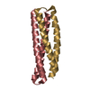
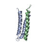
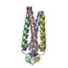
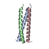
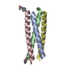
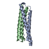
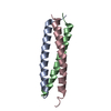
Entry Database : PDB / ID : 1k40Title crystal structure of the FAT domain of focal adhesion kinase adhesion kinase Keywords / Function / homology Function Domain/homology Component
/ / / / / / / / / / / / / / / / / / / / / / / / / / / / / / / / / / / / / / / / / / / / / / / / / / / / / / / / / / / / / / / / / / / / / / / / / / / / / / / / / / / / / / / / / / / / / / / / / / / / / / / / / / / / / / / / / / / / / / / / / / / / / / / / / / / / / / / / / / / / / / / / / / / / / / Biological species Mus musculus (house mouse)Method / / Resolution : 2.25 Å Authors Hayashi, I. / Vuori, K. / Liddington, R.C. Journal : Nat.Struct.Biol. / Year : 2002Title : The focal adhesion targeting (FAT) region of focal adhesion kinase is a four-helix bundle that binds paxillinAuthors : Hayashi, I. / Vuori, K. / Liddington, R.C. History Deposition Oct 4, 2001 Deposition site / Processing site Revision 1.0 Feb 6, 2002 Provider / Type Revision 1.1 Apr 27, 2008 Group Revision 1.2 Jul 13, 2011 Group Revision 1.3 Feb 7, 2024 Group / Database references / Category / chem_comp_bond / database_2Item / _database_2.pdbx_database_accession
Show all Show less
 Movie
Movie Controller


 Open data
 Basic information
 Components
 Keywords
 Function and homology information
 X-RAY DIFFRACTION /
  Authors
 Citation
 Structure visualization
 Downloads & links
 Other downloads
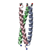


 PDBj
PDBj


 Assembly

 Mass: 18.015 Da / Num. of mol.: 24 / Source method: isolated from a natural source / Formula: H2O
 Sample preparation
 / Beamline: X26C / Wavelength: 1.1 Å
 Processing