 Movie
Movie Controller

[English] 日本語
 Yorodumi
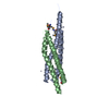
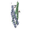
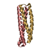
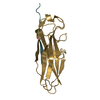
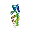
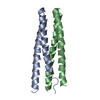
- PDB-3fav: Structure of the CFP10-ESAT6 complex from Mycobacterium tuberculosis -
+ Open data

- Basic information
| Entry | Database: PDB / ID: 3fav | ||||||
|---|---|---|---|---|---|---|---|
| Title | Structure of the CFP10-ESAT6 complex from Mycobacterium tuberculosis | ||||||
 Components Components |
| ||||||
 Keywords Keywords | VIRAL PROTEIN / Complex / Operon structure / Four-helical-bundle / Coiled-coil / WXG-motif / Mycobacterium tuberculosis / secreted / secretion system / adaptor protein / proposed virulent factor | ||||||
| Function / homology |  Function and homology information Function and homology informationsymbiont-mediated suppression of host T-cell mediated immune response / protein secretion by the type VII secretion system / symbiont-mediated perturbation of host signal transduction pathway / Manipulation of host energy metabolism / : / host cell surface binding / host cell endoplasmic reticulum / membrane => GO:0016020 / host cell membrane / peptidoglycan-based cell wall ...symbiont-mediated suppression of host T-cell mediated immune response / protein secretion by the type VII secretion system / symbiont-mediated perturbation of host signal transduction pathway / Manipulation of host energy metabolism / : / host cell surface binding / host cell endoplasmic reticulum / membrane => GO:0016020 / host cell membrane / peptidoglycan-based cell wall / Modulation by Mtb of host immune system / host cell surface / symbiont-mediated suppression of host innate immune response / host cell plasma membrane / protein homodimerization activity / extracellular region / plasma membrane / cytoplasm / cytosol Similarity search - Function | ||||||
| Biological species |   Mycobacterium tuberculosis (bacteria) Mycobacterium tuberculosis (bacteria) | ||||||
| Method |  X-RAY DIFFRACTION / SYNCHROTRON / MOLECULAR REPLACEMENT / Resolution: 2.15 Å X-RAY DIFFRACTION / SYNCHROTRON / MOLECULAR REPLACEMENT / Resolution: 2.15 Å | ||||||
 Authors Authors | Poulsen, C. / Holton, S.J. / Wilmanns, M. / Song, Y.H. | ||||||
 Citation Citation | Journal: Plos One / Year: 2014 Title: WXG100 protein superfamily consists of three subfamilies and exhibits an alpha-helical C-terminal conserved residue pattern. Authors: Poulsen, C. / Panjikar, S. / Holton, S.J. / Wilmanns, M. / Song, Y.H. | ||||||
| History |
|
- Structure visualization
| Structure viewer | Molecule: MolmilJmol/JSmol |
|---|
- Downloads & links
-Download
| PDBx/mmCIF format | 3fav.cif.gz | 135.2 KB | Display | PDBx/mmCIF format |
|---|---|---|---|---|
| PDB format | pdb3fav.ent.gz | 105.1 KB | Display | PDB format |
| PDBx/mmJSON format | 3fav.json.gz | Tree view | PDBx/mmJSON format | |
| Others |  Other downloads Other downloads |
-Validation report
| Arichive directory | https://data.pdbj.org/pub/pdb/validation_reports/fa/3favftp://data.pdbj.org/pub/pdb/validation_reports/fa/3fav | HTTPS FTP |
|---|
-Related structure data
| Related structure data | 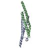 3gvmC  3gwkC 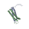 1wa8S S: Starting model for refinement C: citing same article ( |
|---|---|
| Similar structure data |


-Links
 PDBj
PDBj


- Assembly
| Deposited unit | 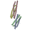
| |||||||||
|---|---|---|---|---|---|---|---|---|---|---|
| 1 | 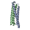
| |||||||||
| 2 | 
| |||||||||
| Unit cell |
| |||||||||
| Components on special symmetry positions |
|
-Components
| #1: Protein | Mass: 10873.819 Da / Num. of mol.: 2 Source method: isolated from a genetically manipulated source Details: The construct contained the entire protein encoding region of the CFP10-ESAT6 operon including its intergenic region Source: (gene. exp.) Mycobacterium tuberculosis (bacteria) / Strain: H37Rv / Gene: esxB / Plasmid details: modified pSD26 vector / Plasmid: pMyNT / Production host: Mycobacterium smegmatis (bacteria) / Strain (production host): MC2 155 / References: UniProt: P0A566, UniProt: P9WNK5*PLUS#2: Protein | Mass: 9777.605 Da / Num. of mol.: 2 Source method: isolated from a genetically manipulated source Details: The construct contained the entire protein encoding region of the CFP10-ESAT6 operon including its intergenic region Source: (gene. exp.) Mycobacterium tuberculosis (bacteria) / Strain: H37Rv / Gene: esxA / Plasmid details: modified pSD26 vector / Plasmid: pMyNT / Production host: Mycobacterium smegmatis (bacteria) / Strain (production host): MC2 155 / References: UniProt: P0A564, UniProt: P9WNK7*PLUS#3: Chemical | ChemComp-ZN /   Mass: 65.409 Da / Num. of mol.: 7 / Source method: obtained synthetically / Formula: Zn Mass: 65.409 Da / Num. of mol.: 7 / Source method: obtained synthetically / Formula: Zn#4: Chemical | ChemComp-IMD / |   Mass: 69.085 Da / Num. of mol.: 1 / Source method: obtained synthetically / Formula: C3H5N2 Mass: 69.085 Da / Num. of mol.: 1 / Source method: obtained synthetically / Formula: C3H5N2#5: Water | ChemComp-HOH / |  Mass: 18.015 Da / Num. of mol.: 308 / Source method: isolated from a natural source / Formula: H2O Mass: 18.015 Da / Num. of mol.: 308 / Source method: isolated from a natural source / Formula: H2O |
|---|
-Experimental details
-Experiment
| Experiment | Method: X-RAY DIFFRACTION / Number of used crystals: 1 |
|---|
- Sample preparation
| Crystal | Density Matthews: 1.94 Å3/Da / Density % sol: 36.66 % |
|---|---|
| Crystal grow | Temperature: 292 K / Method: vapor diffusion, hanging drop / pH: 6.5 Details: 33% (v/v) PEG 600, 200mM zinc acetate, 100mM imidazol pH 6.5, VAPOR DIFFUSION, HANGING DROP, temperature 292K |
-Data collection
| Diffraction | Mean temperature: 100 K |
|---|---|
| Diffraction source | Source: SYNCHROTRON / Site: ESRF  / Beamline: BM14 / Wavelength: 1.033 Å / Beamline: BM14 / Wavelength: 1.033 Å |
| Detector | Type: MARMOSAIC 225 mm CCD / Detector: CCD / Date: Nov 11, 2006 |
| Radiation | Monochromator: Si(111) / Protocol: SINGLE WAVELENGTH / Monochromatic (M) / Laue (L): M / Scattering type: x-ray |
| Radiation wavelength | Wavelength: 1.033 Å / Relative weight: 1 |
| Reflection | Resolution: 2.15→20 Å / Num. all: 18161 / Num. obs: 17336 / % possible obs: 95.5 % / Observed criterion σ(I): -3 / Redundancy: 3.35 % / Biso Wilson estimate: 30.407 Å2 / Rmerge(I) obs: 0.055 / Rsym value: 0.065 / Net I/σ(I): 14.4 |
| Reflection shell | Resolution: 2.15→2.21 Å / Redundancy: 2.2 % / Rmerge(I) obs: 0.197 / Mean I/σ(I) obs: 4.2 / Num. measured obs: 2149 / Num. unique all: 1333 / Num. unique obs: 978 / Rsym value: 0.261 / % possible all: 73.4 |
- Processing
| Software |
| ||||||||||||||||||||||||||||||||||||||||||||||||||||||||||||||||||||||||||||||||||||||||||||||||||||||||||||||||||||||||||||||||||||||||||||||||||||||||||||||||||||||||||||||||||||||||||||||||||||||
|---|---|---|---|---|---|---|---|---|---|---|---|---|---|---|---|---|---|---|---|---|---|---|---|---|---|---|---|---|---|---|---|---|---|---|---|---|---|---|---|---|---|---|---|---|---|---|---|---|---|---|---|---|---|---|---|---|---|---|---|---|---|---|---|---|---|---|---|---|---|---|---|---|---|---|---|---|---|---|---|---|---|---|---|---|---|---|---|---|---|---|---|---|---|---|---|---|---|---|---|---|---|---|---|---|---|---|---|---|---|---|---|---|---|---|---|---|---|---|---|---|---|---|---|---|---|---|---|---|---|---|---|---|---|---|---|---|---|---|---|---|---|---|---|---|---|---|---|---|---|---|---|---|---|---|---|---|---|---|---|---|---|---|---|---|---|---|---|---|---|---|---|---|---|---|---|---|---|---|---|---|---|---|---|---|---|---|---|---|---|---|---|---|---|---|---|---|---|---|---|
| Refinement | Method to determine structure: MOLECULAR REPLACEMENT Starting model: PDB ENTRY 1WA8 Resolution: 2.15→19.858 Å / Occupancy max: 1 / Occupancy min: 0 / FOM work R set: 0.847 / Cross valid method: THROUGHOUT / Stereochemistry target values: ML
| ||||||||||||||||||||||||||||||||||||||||||||||||||||||||||||||||||||||||||||||||||||||||||||||||||||||||||||||||||||||||||||||||||||||||||||||||||||||||||||||||||||||||||||||||||||||||||||||||||||||
| Solvent computation | Bsol: 61.173 Å2 / ksol: 0.39 e/Å3 | ||||||||||||||||||||||||||||||||||||||||||||||||||||||||||||||||||||||||||||||||||||||||||||||||||||||||||||||||||||||||||||||||||||||||||||||||||||||||||||||||||||||||||||||||||||||||||||||||||||||
| Displacement parameters | Biso max: 166.54 Å2 / Biso mean: 30.573 Å2 / Biso min: 11.89 Å2
| ||||||||||||||||||||||||||||||||||||||||||||||||||||||||||||||||||||||||||||||||||||||||||||||||||||||||||||||||||||||||||||||||||||||||||||||||||||||||||||||||||||||||||||||||||||||||||||||||||||||
| Refinement step | Cycle: LAST / Resolution: 2.15→19.858 Å
| ||||||||||||||||||||||||||||||||||||||||||||||||||||||||||||||||||||||||||||||||||||||||||||||||||||||||||||||||||||||||||||||||||||||||||||||||||||||||||||||||||||||||||||||||||||||||||||||||||||||
| Refine LS restraints |
| ||||||||||||||||||||||||||||||||||||||||||||||||||||||||||||||||||||||||||||||||||||||||||||||||||||||||||||||||||||||||||||||||||||||||||||||||||||||||||||||||||||||||||||||||||||||||||||||||||||||
| LS refinement shell |
| ||||||||||||||||||||||||||||||||||||||||||||||||||||||||||||||||||||||||||||||||||||||||||||||||||||||||||||||||||||||||||||||||||||||||||||||||||||||||||||||||||||||||||||||||||||||||||||||||||||||
| Refinement TLS params. | S33: 0 Å ° / Method: refined / Refine-ID: X-RAY DIFFRACTION
| ||||||||||||||||||||||||||||||||||||||||||||||||||||||||||||||||||||||||||||||||||||||||||||||||||||||||||||||||||||||||||||||||||||||||||||||||||||||||||||||||||||||||||||||||||||||||||||||||||||||
| Refinement TLS group |
|
