 Movie
Movie Controller

[English] 日本語
 Yorodumi
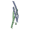
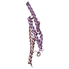
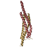
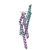
- PDB-3gvm: Structure of the homodimeric WXG-100 family protein from Streptoc... -
+ Open data

- Basic information
| Entry | Database: PDB / ID: 3gvm | ||||||
|---|---|---|---|---|---|---|---|
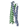
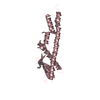
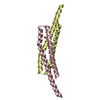
| Title | Structure of the homodimeric WXG-100 family protein from Streptococcus agalactiae | ||||||
 Components Components | Putative uncharacterized protein SAG1039 | ||||||
 Keywords Keywords | VIRAL PROTEIN / WXG motif / Four-helical bundle | ||||||
| Function / homology | ESAT-6-like superfamily / Type VII secretion system ESAT-6-like / Proteins of 100 residues with WXG / ESAT-6-like / Helix Hairpins / Orthogonal Bundle / Mainly Alpha / ESAT-6-like protein SAG1039 Function and homology information Function and homology information | ||||||
| Biological species |  Streptococcus agalactiae serogroup V (bacteria) Streptococcus agalactiae serogroup V (bacteria) | ||||||
| Method |  X-RAY DIFFRACTION / SYNCHROTRON / SAD / Resolution: 2.15 Å X-RAY DIFFRACTION / SYNCHROTRON / SAD / Resolution: 2.15 Å | ||||||
 Authors Authors | Poulsen, C. / Gries, F. / Wilmanns, M. / Song, Y.H. | ||||||
 Citation Citation | Journal: Plos One / Year: 2014 Title: WXG100 protein superfamily consists of three subfamilies and exhibits an alpha-helical C-terminal conserved residue pattern. Authors: Poulsen, C. / Panjikar, S. / Holton, S.J. / Wilmanns, M. / Song, Y.H. | ||||||
| History |
|
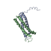
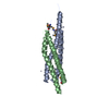
- Structure visualization
| Structure viewer | Molecule: MolmilJmol/JSmol |
|---|
- Downloads & links
-Download
| PDBx/mmCIF format | 3gvm.cif.gz | 93.9 KB | Display | PDBx/mmCIF format |
|---|---|---|---|---|
| PDB format | pdb3gvm.ent.gz | 72.7 KB | Display | PDB format |
| PDBx/mmJSON format | 3gvm.json.gz | Tree view | PDBx/mmJSON format | |
| Others |  Other downloads Other downloads |
-Validation report
| Arichive directory | https://data.pdbj.org/pub/pdb/validation_reports/gv/3gvmftp://data.pdbj.org/pub/pdb/validation_reports/gv/3gvm | HTTPS FTP |
|---|
-Related structure data


-Links
 PDBj
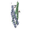
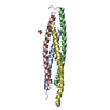
PDBj- Assembly
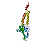
| Deposited unit | 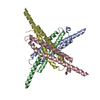
| ||||||||
|---|---|---|---|---|---|---|---|---|---|
| 1 | 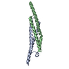
| ||||||||
| 2 | 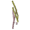
| ||||||||
| Unit cell |
|
-Components
| #1: Protein | Mass: 10838.833 Da / Num. of mol.: 4 Source method: isolated from a genetically manipulated source Source: (gene. exp.) Streptococcus agalactiae serogroup V (bacteria)Strain: 2603V/R / Gene: SAG1039 / Plasmid: pETM11 / Production host: #2: Water | ChemComp-HOH / |  Mass: 18.015 Da / Num. of mol.: 462 / Source method: isolated from a natural source / Formula: H2O Mass: 18.015 Da / Num. of mol.: 462 / Source method: isolated from a natural source / Formula: H2O |
|---|
-Experimental details
-Experiment
| Experiment | Method: X-RAY DIFFRACTION / Number of used crystals: 2 |
|---|
- Sample preparation
| Crystal | Density Matthews: 4.44 Å3/Da / Density % sol: 72.31 % |
|---|---|
| Crystal grow | Temperature: 297 K / Method: vapor diffusion, hanging drop / pH: 8 Details: 0.1M Tris-HCl pH 8.0, 1.9M (NH4)SO4, VAPOR DIFFUSION, HANGING DROP, temperature 297.0K |
-Data collection
| Diffraction |
| ||||||||||||||||||||||||||||||||||||||||||||||||||||||||||||||||||||||||||||||||||||||||||||||||||||||||||||||||||||||||||||||
|---|---|---|---|---|---|---|---|---|---|---|---|---|---|---|---|---|---|---|---|---|---|---|---|---|---|---|---|---|---|---|---|---|---|---|---|---|---|---|---|---|---|---|---|---|---|---|---|---|---|---|---|---|---|---|---|---|---|---|---|---|---|---|---|---|---|---|---|---|---|---|---|---|---|---|---|---|---|---|---|---|---|---|---|---|---|---|---|---|---|---|---|---|---|---|---|---|---|---|---|---|---|---|---|---|---|---|---|---|---|---|---|---|---|---|---|---|---|---|---|---|---|---|---|---|---|---|---|
| Diffraction source |
| ||||||||||||||||||||||||||||||||||||||||||||||||||||||||||||||||||||||||||||||||||||||||||||||||||||||||||||||||||||||||||||||
| Detector |
| ||||||||||||||||||||||||||||||||||||||||||||||||||||||||||||||||||||||||||||||||||||||||||||||||||||||||||||||||||||||||||||||
| Radiation |
| ||||||||||||||||||||||||||||||||||||||||||||||||||||||||||||||||||||||||||||||||||||||||||||||||||||||||||||||||||||||||||||||
| Radiation wavelength |
| ||||||||||||||||||||||||||||||||||||||||||||||||||||||||||||||||||||||||||||||||||||||||||||||||||||||||||||||||||||||||||||||
| Reflection | Number: 138669 / Rmerge(I) obs: 0.04 / D res high: 2.5 Å / Num. obs: 50045 / % possible obs: 98 | ||||||||||||||||||||||||||||||||||||||||||||||||||||||||||||||||||||||||||||||||||||||||||||||||||||||||||||||||||||||||||||||
| Diffraction reflection shell |
| ||||||||||||||||||||||||||||||||||||||||||||||||||||||||||||||||||||||||||||||||||||||||||||||||||||||||||||||||||||||||||||||
| Reflection | Resolution: 2.15→50 Å / Num. all: 43035 / Num. obs: 43011 / % possible obs: 99.9 % / Observed criterion σ(I): -3 / Redundancy: 6.1 % / Biso Wilson estimate: 31.776 Å2 / Rmerge(I) obs: 0.091 | ||||||||||||||||||||||||||||||||||||||||||||||||||||||||||||||||||||||||||||||||||||||||||||||||||||||||||||||||||||||||||||||
| Reflection shell | Resolution: 2.15→2.21 Å / Redundancy: 6.2 % / Rmerge(I) obs: 0.507 / Mean I/σ(I) obs: 3.8 / Num. measured obs: 19300 / Num. unique all: 3130 / Num. unique obs: 3128 / % possible all: 99.9 |


- Processing
| Software |
| ||||||||||||||||||||||||||||||||||||||||||||||||||||||||||||||||||||||||||||||||||||||||||||||||||||||||||||||||
|---|---|---|---|---|---|---|---|---|---|---|---|---|---|---|---|---|---|---|---|---|---|---|---|---|---|---|---|---|---|---|---|---|---|---|---|---|---|---|---|---|---|---|---|---|---|---|---|---|---|---|---|---|---|---|---|---|---|---|---|---|---|---|---|---|---|---|---|---|---|---|---|---|---|---|---|---|---|---|---|---|---|---|---|---|---|---|---|---|---|---|---|---|---|---|---|---|---|---|---|---|---|---|---|---|---|---|---|---|---|---|---|---|---|
| Refinement | Method to determine structure: SAD / Resolution: 2.15→46.957 Å / Occupancy max: 1 / Occupancy min: 0.36 / FOM work R set: 0.871 / SU ML: -0 / σ(F): 2 / Stereochemistry target values: ML
| ||||||||||||||||||||||||||||||||||||||||||||||||||||||||||||||||||||||||||||||||||||||||||||||||||||||||||||||||
| Solvent computation | Shrinkage radii: 0.9 Å / VDW probe radii: 1.11 Å / Solvent model: FLAT BULK SOLVENT MODEL / Bsol: 57.188 Å2 / ksol: 0.369 e/Å3 | ||||||||||||||||||||||||||||||||||||||||||||||||||||||||||||||||||||||||||||||||||||||||||||||||||||||||||||||||
| Displacement parameters | Biso max: 94.94 Å2 / Biso mean: 30.376 Å2 / Biso min: 12.8 Å2
| ||||||||||||||||||||||||||||||||||||||||||||||||||||||||||||||||||||||||||||||||||||||||||||||||||||||||||||||||
| Refinement step | Cycle: LAST / Resolution: 2.15→46.957 Å
| ||||||||||||||||||||||||||||||||||||||||||||||||||||||||||||||||||||||||||||||||||||||||||||||||||||||||||||||||
| Refine LS restraints |
| ||||||||||||||||||||||||||||||||||||||||||||||||||||||||||||||||||||||||||||||||||||||||||||||||||||||||||||||||
| LS refinement shell | Refine-ID: X-RAY DIFFRACTION / Total num. of bins used: 15
|
