 Movie
Movie Controller

[English] 日本語
 Yorodumi
- PDB-1gzl: Crystal structure of C14linkmid/IQN17: a cross-linked inhibitor o... -
+ Open data

- Basic information
| Entry | Database: PDB / ID: 1gzl | ||||||
|---|---|---|---|---|---|---|---|
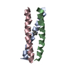
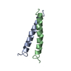
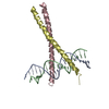
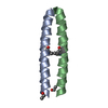
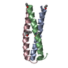
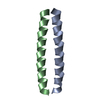
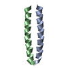
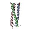
| Title | Crystal structure of C14linkmid/IQN17: a cross-linked inhibitor of HIV-1 entry bound to the gp41 hydrophobic pocket | ||||||
 Components Components |
| ||||||
 Keywords Keywords | GLYCOPROTEIN / HIV ENTRY / INHIBITOR / CROSS-LINK / GP41 / COILED COIL | ||||||
| Function / homology |  Function and homology information Function and homology informationSynthesis and processing of ENV and VPU / FCERI mediated MAPK activation / protein localization to nuclear periphery / Activation of the AP-1 family of transcription factors / negative regulation of ribosomal protein gene transcription by RNA polymerase II / symbiont-mediated evasion of host immune response / positive regulation of cellular response to amino acid starvation / response to amino acid starvation / mediator complex binding / positive regulation of establishment of T cell polarity ...Synthesis and processing of ENV and VPU / FCERI mediated MAPK activation / protein localization to nuclear periphery / Activation of the AP-1 family of transcription factors / negative regulation of ribosomal protein gene transcription by RNA polymerase II / symbiont-mediated evasion of host immune response / positive regulation of cellular response to amino acid starvation / response to amino acid starvation / mediator complex binding / positive regulation of establishment of T cell polarity / Alpha-defensins / Oxidative Stress Induced Senescence / Dectin-2 family / amino acid biosynthetic process / TFIID-class transcription factor complex binding / Binding and entry of HIV virion / positive regulation of RNA polymerase II transcription preinitiation complex assembly / positive regulation of transcription initiation by RNA polymerase II / positive regulation of plasma membrane raft polarization / positive regulation of receptor clustering / cellular response to nutrient levels / host cell endosome membrane / cellular response to amino acid starvation / actin filament organization / Assembly Of The HIV Virion / Budding and maturation of HIV virion / RNA polymerase II transcription regulator complex / DNA-binding transcription activator activity, RNA polymerase II-specific / transcription regulator complex / clathrin-dependent endocytosis of virus by host cell / sequence-specific DNA binding / RNA polymerase II-specific DNA-binding transcription factor binding / DNA-binding transcription factor activity, RNA polymerase II-specific / intracellular signal transduction / viral protein processing / RNA polymerase II cis-regulatory region sequence-specific DNA binding / DNA-binding transcription factor activity / receptor ligand activity / fusion of virus membrane with host plasma membrane / fusion of virus membrane with host endosome membrane / viral envelope / chromatin binding / symbiont entry into host cell / virion attachment to host cell / host cell plasma membrane / virion membrane / structural molecule activity / negative regulation of transcription by RNA polymerase II / positive regulation of transcription by RNA polymerase II / membrane / identical protein binding / nucleus Similarity search - Function | ||||||
| Biological species |    HUMAN IMMUNODEFICIENCY VIRUS 1HUMAN IMMUNODEFICIENCY VIRUS HUMAN IMMUNODEFICIENCY VIRUS 1HUMAN IMMUNODEFICIENCY VIRUS | ||||||
| Method |  X-RAY DIFFRACTION / SYNCHROTRON / MOLECULAR REPLACEMENT / Resolution: 1.8 Å X-RAY DIFFRACTION / SYNCHROTRON / MOLECULAR REPLACEMENT / Resolution: 1.8 Å | ||||||
 Authors Authors | Sia, S.K. / Carr, P.A. / Cochran, A.G. / Malashkevich, V.M. / Kim, P.S. | ||||||
 Citation Citation | Journal: Proc.Natl.Acad.Sci.USA / Year: 2002 Title: Short Constrained Peptides that Inhibit HIV-1 Entry Authors: Sia, S.K. / Carr, P.A. / Cochran, A.G. / Malashkevich, V.M. / Kim, P.S. | ||||||
| History |
|
- Structure visualization
| Structure viewer | Molecule: MolmilJmol/JSmol |
|---|
- Downloads & links
-Download
| PDBx/mmCIF format | 1gzl.cif.gz | 40.2 KB | Display | PDBx/mmCIF format |
|---|---|---|---|---|
| PDB format | pdb1gzl.ent.gz | 28.7 KB | Display | PDB format |
| PDBx/mmJSON format | 1gzl.json.gz | Tree view | PDBx/mmJSON format | |
| Others |  Other downloads Other downloads |
-Validation report
| Arichive directory | https://data.pdbj.org/pub/pdb/validation_reports/gz/1gzlftp://data.pdbj.org/pub/pdb/validation_reports/gz/1gzl | HTTPS FTP |
|---|
-Related structure data
| Related structure data | |
|---|---|
| Similar structure data |


-Links
 PDBj
PDBj


- Assembly
| Deposited unit | 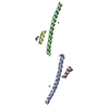
| |||||||||
|---|---|---|---|---|---|---|---|---|---|---|
| 1 | 
| |||||||||
| 2 | 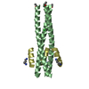
| |||||||||
| Unit cell |
| |||||||||
| Components on special symmetry positions |
| |||||||||
| Details | AND CHAINS B AND D). APPLYING CRYSTAL SYMMETRY GENERATES TWO TRIMERS OF HETERODIMERS (HEXAMERS). |
-Components
| #1: Protein/peptide | Mass: 5468.566 Da / Num. of mol.: 2 Fragment: GP41 HYDROPHOBIC POCKET, RESIDUES 565-581, GCN4, RESIDUES 249-276 Source method: obtained synthetically / Details: THIS PEPTIDE WAS CHEMICALLY SYNTHESIZED Source: (synth.) HUMAN IMMUNODEFICIENCY VIRUS 1References: UniProt: P03069, UniProt: P04578 #2: Protein/peptide | Mass: 1670.710 Da / Num. of mol.: 2 / Fragment: RESIDUES 628-639 / Mutation: YES / Source method: obtained synthetically / Details: THIS PEPTIDE WAS CHEMICALLY SYNTHESIZED / Source: (synth.) HUMAN IMMUNODEFICIENCY VIRUS / References: UniProt: P04578#3: Chemical |   Mass: 35.453 Da / Num. of mol.: 2 / Source method: obtained synthetically / Formula: Cl Mass: 35.453 Da / Num. of mol.: 2 / Source method: obtained synthetically / Formula: Cl#4: Chemical |   Mass: 102.178 Da / Num. of mol.: 2 / Source method: obtained synthetically / Formula: C5H14N2 Mass: 102.178 Da / Num. of mol.: 2 / Source method: obtained synthetically / Formula: C5H14N2#5: Water | ChemComp-HOH / |  Mass: 18.015 Da / Num. of mol.: 77 / Source method: isolated from a natural source / Formula: H2O Mass: 18.015 Da / Num. of mol.: 77 / Source method: isolated from a natural source / Formula: H2OCompound details | ENGINEERED | Has protein modification | Y | Sequence details | IN CHAINS C AND D, MET 629 AND ASN 636 ARE MUTATED TO GLUTAMIC ACID. A DIAMINOPENTANE GROUP LINKS ...IN CHAINS C AND D, MET 629 AND ASN 636 ARE MUTATED TO GLUTAMIC ACID. A DIAMINOPEN | |
|---|
-Experimental details
-Experiment
| Experiment | Method: X-RAY DIFFRACTION / Number of used crystals: 1 |
|---|
- Sample preparation
| Crystal | Density Matthews: 2.46 Å3/Da / Density % sol: 49.6 % | ||||||||||||||||||||||||||||||
|---|---|---|---|---|---|---|---|---|---|---|---|---|---|---|---|---|---|---|---|---|---|---|---|---|---|---|---|---|---|---|---|
| Crystal grow | pH: 8.6 / Details: 16% ISOPROPANOL, 0.1 M TRIS, PH 8.6, 1 M (NH4)2SO4 | ||||||||||||||||||||||||||||||
| Crystal grow | *PLUS Method: vapor diffusion, hanging drop | ||||||||||||||||||||||||||||||
| Components of the solutions | *PLUS
|
-Data collection
| Diffraction | Mean temperature: 100 K |
|---|---|
| Diffraction source | Source: SYNCHROTRON / Site: NSLS  / Beamline: X4A / Wavelength: 1.0093 / Beamline: X4A / Wavelength: 1.0093 |
| Detector | Type: ADSC CCD / Detector: CCD / Date: Jun 19, 2000 |
| Radiation | Monochromator: GRAPHITE / Protocol: SINGLE WAVELENGTH / Monochromatic (M) / Laue (L): M / Scattering type: x-ray |
| Radiation wavelength | Wavelength: 1.0093 Å / Relative weight: 1 |
| Reflection | Resolution: 1.8→20 Å / Num. obs: 59821 / % possible obs: 91.9 % / Observed criterion σ(I): 0 / Redundancy: 5 % / Biso Wilson estimate: 26.9 Å2 / Rsym value: 0.044 / Net I/σ(I): 13.6 |
| Reflection shell | Resolution: 1.8→1.86 Å / Redundancy: 2.1 % / Rmerge(I) obs: 0.187 / Mean I/σ(I) obs: 7.2 / % possible all: 69.5 |
| Reflection | *PLUS Highest resolution: 1.86 Å / Num. obs: 11190 / % possible obs: 94.3 % / Num. measured all: 59821 / Rmerge(I) obs: 0.044 |
| Reflection shell | *PLUS % possible obs: 80.2 % / Rmerge(I) obs: 0.15 |
- Processing
| Software |
| ||||||||||||||||||||||||||||||||||||||||||||||||||||||||||||||||||||||||||||||||
|---|---|---|---|---|---|---|---|---|---|---|---|---|---|---|---|---|---|---|---|---|---|---|---|---|---|---|---|---|---|---|---|---|---|---|---|---|---|---|---|---|---|---|---|---|---|---|---|---|---|---|---|---|---|---|---|---|---|---|---|---|---|---|---|---|---|---|---|---|---|---|---|---|---|---|---|---|---|---|---|---|---|
| Refinement | Method to determine structure: MOLECULAR REPLACEMENT Starting model: UNBOUND IQN17 AND A MODEL OF C14LINKMID BOUND TO THE HYDROPHOBIC POCKET Resolution: 1.8→19.58 Å / Rfactor Rfree error: 0.009 / Data cutoff high absF: 505253.07 / Isotropic thermal model: RESTRAINED / Cross valid method: THROUGHOUT / σ(F): 0 Details: FINAL REFINEMENT IS AFTER DETWINNING DATA. 529 REFLECTIONS WERE REJECTED BY CNS AFTER MEROHEDRAL DETWINNING. THE THREE CHAINS OF THE TRIMER ARE RELATED BY CRYSTALLOGRAPHIC SYMMETRY. TO ...Details: FINAL REFINEMENT IS AFTER DETWINNING DATA. 529 REFLECTIONS WERE REJECTED BY CNS AFTER MEROHEDRAL DETWINNING. THE THREE CHAINS OF THE TRIMER ARE RELATED BY CRYSTALLOGRAPHIC SYMMETRY. TO GENERATE THE TRIMER, APPLY SYMMETRY TRANSFORMATIONS. THE FINAL MODEL CONSISTS OF ALL RESIDUES EXCEPT FOR THE TWO N-TERMINAL RESIDUES OF C14LINKMID, WHICH ARE DISORDERED.
| ||||||||||||||||||||||||||||||||||||||||||||||||||||||||||||||||||||||||||||||||
| Solvent computation | Solvent model: FLAT MODEL / Bsol: 97.565 Å2 / ksol: 0.428224 e/Å3 | ||||||||||||||||||||||||||||||||||||||||||||||||||||||||||||||||||||||||||||||||
| Displacement parameters | Biso mean: 36 Å2
| ||||||||||||||||||||||||||||||||||||||||||||||||||||||||||||||||||||||||||||||||
| Refine analyze |
| ||||||||||||||||||||||||||||||||||||||||||||||||||||||||||||||||||||||||||||||||
| Refinement step | Cycle: LAST / Resolution: 1.8→19.58 Å
| ||||||||||||||||||||||||||||||||||||||||||||||||||||||||||||||||||||||||||||||||
| Refine LS restraints |
| ||||||||||||||||||||||||||||||||||||||||||||||||||||||||||||||||||||||||||||||||
| LS refinement shell | Resolution: 1.8→1.91 Å / Rfactor Rfree error: 0.034 / Total num. of bins used: 6
| ||||||||||||||||||||||||||||||||||||||||||||||||||||||||||||||||||||||||||||||||
| Xplor file |
| ||||||||||||||||||||||||||||||||||||||||||||||||||||||||||||||||||||||||||||||||
| Refinement | *PLUS Highest resolution: 1.86 Å / Lowest resolution: 20 Å / Num. reflection obs: 10090 / Num. reflection Rfree: 687 | ||||||||||||||||||||||||||||||||||||||||||||||||||||||||||||||||||||||||||||||||
| Solvent computation | *PLUS | ||||||||||||||||||||||||||||||||||||||||||||||||||||||||||||||||||||||||||||||||
| Displacement parameters | *PLUS | ||||||||||||||||||||||||||||||||||||||||||||||||||||||||||||||||||||||||||||||||
| Refine LS restraints | *PLUS
| ||||||||||||||||||||||||||||||||||||||||||||||||||||||||||||||||||||||||||||||||
| LS refinement shell | *PLUS Rfactor Rfree: 0.271 / Rfactor Rwork: 0.297 |
