 Movie
Movie Controller

[English] 日本語
 Yorodumi
- PDB-1gk6: Human vimentin coil 2B fragment linked to GCN4 leucine zipper (Z2B) -
+ Open data

- Basic information
| Entry | Database: PDB / ID: 1gk6 | |||||||||
|---|---|---|---|---|---|---|---|---|---|---|
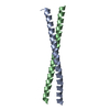
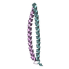
| Title | Human vimentin coil 2B fragment linked to GCN4 leucine zipper (Z2B) | |||||||||
 Components Components | VIMENTIN | |||||||||
 Keywords Keywords | STRUCTURAL PROTEIN / INTERMEDIATE FILAMENT / DIMER / PARALLEL COILED COIL / HEPTAD REPEAT / LEUCINE ZIPPER / FUSION PROTEIN | |||||||||
| Function / homology |  Function and homology information Function and homology informationkeratin filament binding / lens fiber cell development / intermediate filament organization / cellular response to muramyl dipeptide / structural constituent of eye lens / FCERI mediated MAPK activation / protein localization to nuclear periphery / Activation of the AP-1 family of transcription factors / negative regulation of ribosomal protein gene transcription by RNA polymerase II / positive regulation of cellular response to amino acid starvation ...keratin filament binding / lens fiber cell development / intermediate filament organization / cellular response to muramyl dipeptide / structural constituent of eye lens / FCERI mediated MAPK activation / protein localization to nuclear periphery / Activation of the AP-1 family of transcription factors / negative regulation of ribosomal protein gene transcription by RNA polymerase II / positive regulation of cellular response to amino acid starvation / response to amino acid starvation / mediator complex binding / astrocyte development / intermediate filament cytoskeleton / Oxidative Stress Induced Senescence / Striated Muscle Contraction / RHOBTB1 GTPase cycle / intermediate filament / Bergmann glial cell differentiation / Developmental Lineage of Mammary Gland Myoepithelial Cells / cell leading edge / positive regulation of collagen biosynthetic process / amino acid biosynthetic process / TFIID-class transcription factor complex binding / microtubule organizing center / positive regulation of RNA polymerase II transcription preinitiation complex assembly / Caspase-mediated cleavage of cytoskeletal proteins / positive regulation of transcription initiation by RNA polymerase II / phagocytic vesicle / regulation of mRNA stability / cellular response to nutrient levels / cellular response to amino acid starvation / Late endosomal microautophagy / structural constituent of cytoskeleton / cellular response to type II interferon / RNA polymerase II transcription regulator complex / nuclear matrix / Chaperone Mediated Autophagy / neuron projection development / Aggrephagy / peroxisome / negative regulation of neuron projection development / double-stranded RNA binding / cellular response to lipopolysaccharide / Interleukin-4 and Interleukin-13 signaling / scaffold protein binding / DNA-binding transcription activator activity, RNA polymerase II-specific / transcription regulator complex / sequence-specific DNA binding / molecular adaptor activity / cytoskeleton / RNA polymerase II-specific DNA-binding transcription factor binding / DNA-binding transcription factor activity, RNA polymerase II-specific / intracellular signal transduction / RNA polymerase II cis-regulatory region sequence-specific DNA binding / DNA-binding transcription factor activity / protein domain specific binding / axon / focal adhesion / chromatin binding / positive regulation of gene expression / negative regulation of transcription by RNA polymerase II / positive regulation of transcription by RNA polymerase II / extracellular exosome / identical protein binding / nucleus / plasma membrane / cytoplasm / cytosol Similarity search - Function | |||||||||
| Biological species |   HOMO SAPIENS (human) HOMO SAPIENS (human) | |||||||||
| Method |  X-RAY DIFFRACTION / SYNCHROTRON / MOLECULAR REPLACEMENT / Resolution: 1.9 Å X-RAY DIFFRACTION / SYNCHROTRON / MOLECULAR REPLACEMENT / Resolution: 1.9 Å | |||||||||
 Authors Authors | Strelkov, S.V. / Herrmann, H. / Geisler, N. / Zimbelmann, R. / Aebi, U. / Burkhard, P. | |||||||||
 Citation Citation | Journal: Embo J. / Year: 2002 Title: Conserved Segments 1A and 2B of the Intermediate Filament Dimer: Their Atomic Structures and Role in Filament Assembly. Authors: Strelkov, S. / Herrmann, H. / Geisler, N. / Wedig, T. / Zimbelmann, R. / Aebi, U. / Burkhard, P. #1: Journal: J.Mol.Biol. / Year: 2001 Title: Divide-and-Conquer Crystallographic Approach Towards an Atomic Structure of Intermediate Filaments Authors: Strelkov, S.V. / Herrmann, H. / Geisler, N. / Lustig, A. / Ivaninskii, S. / Zimbelmann, R. / Burkhard, P. / Aebi, U. #2: Journal: J. Mol. Biol. / Year: 2000 Title: The Intermediate Filament Protein Consensus Motifof Helix 2B: Its Atomic Structure and Contribution to Assembly Authors: Herrmann, H. / Strelkov, S.V. / Feja, B. / Rogers, K.R. / Brettel, M. / Lustig, A. / Haener, M. / Parry, D.A.D. / Steinert, P.M. / Burkhard, P. / Aebi, U. | |||||||||
| History |
|
- Structure visualization
| Structure viewer | Molecule: MolmilJmol/JSmol |
|---|
- Downloads & links
-Download
| PDBx/mmCIF format | 1gk6.cif.gz | 37.4 KB | Display | PDBx/mmCIF format |
|---|---|---|---|---|
| PDB format | pdb1gk6.ent.gz | 27.2 KB | Display | PDB format |
| PDBx/mmJSON format | 1gk6.json.gz | Tree view | PDBx/mmJSON format | |
| Others |  Other downloads Other downloads |
-Validation report
| Arichive directory | https://data.pdbj.org/pub/pdb/validation_reports/gk/1gk6ftp://data.pdbj.org/pub/pdb/validation_reports/gk/1gk6 | HTTPS FTP |
|---|
-Related structure data
| Related structure data |  1gk4C 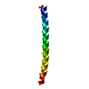 1gk7C 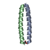 2ztaS C: citing same article ( S: Starting model for refinement |
|---|---|
| Similar structure data |


-Links
 PDBj
PDBj


- Assembly
| Deposited unit | 
| ||||||||
|---|---|---|---|---|---|---|---|---|---|
| 1 |
| ||||||||
| Unit cell |
|
-Components
| #1: Protein | Mass: 6926.064 Da / Num. of mol.: 2 Fragment: Z2B FUSION CONSTRUCT CONTAINING THE GCN4 LEUCINE ZIPPER LINKED TO VIMENTIN RESIDUES 385 - 412 Source method: isolated from a genetically manipulated source Details: N-TERMINAL HALF OF THE MOLECULE CONTAINS THE GCN4 LEUCINE ZIPPER SEQUENCE WHILE THE C-TERMINAL HALF CONTAINS THE VIMENTIN SEQUENCE Source: (gene. exp.) HOMO SAPIENS (human)Production host:  #2: Water | ChemComp-HOH / |  Mass: 18.015 Da / Num. of mol.: 194 / Source method: isolated from a natural source / Formula: H2O Mass: 18.015 Da / Num. of mol.: 194 / Source method: isolated from a natural source / Formula: H2OSequence details | THE RESIDUE NUMBERING USED IN THE LITERATURE FOR VIMENTIN DIFFERS BY +1 FROM THE NUMBERING USED IN ...THE RESIDUE NUMBERING USED IN THE LITERATURE | |
|---|
-Experimental details
-Experiment
| Experiment | Method: X-RAY DIFFRACTION / Number of used crystals: 1 |
|---|
- Sample preparation
| Crystal | Density Matthews: 3.72 Å3/Da / Density % sol: 67.5 % |
|---|---|
| Crystal grow | Method: vapor diffusion, hanging drop / pH: 8.5 Details: HANGING DROPS WITH 12.5MG/ML PROTEIN AND 0.55M (NH4)2HPO4, PH ADJUSTED TO 9.0 WITH NAOH, AS PRECIPITANT |
| Crystal grow | *PLUS Temperature: 20 ℃ / Method: vapor diffusion, hanging drop / Details: Strelkov, S.V., (2001) J.Mol.Biol., 306, 773. |
| Components of the solutions | *PLUS Conc.: 10-15 mg/ml / Common name: protein |
-Data collection
| Diffraction | Mean temperature: 100 K |
|---|---|
| Diffraction source | Source: SYNCHROTRON / Site: EMBL/DESY, HAMBURG  / Beamline: X31 / Wavelength: 1.2545 / Beamline: X31 / Wavelength: 1.2545 |
| Detector | Type: MARRESEARCH / Detector: IMAGE PLATE / Date: Nov 20, 1998 |
| Radiation | Protocol: SINGLE WAVELENGTH / Monochromatic (M) / Laue (L): M / Scattering type: x-ray |
| Radiation wavelength | Wavelength: 1.2545 Å / Relative weight: 1 |
| Reflection | Resolution: 1.9→33.5 Å / Num. obs: 16241 / % possible obs: 99.2 % / Observed criterion σ(I): 0 / Redundancy: 3.7 % / Rmerge(I) obs: 0.063 / Net I/σ(I): 15 |
| Reflection shell | Resolution: 1.9→1.97 Å / Redundancy: 3.6 % / Rmerge(I) obs: 0.372 / Mean I/σ(I) obs: 3.3 / % possible all: 99.4 |
| Reflection | *PLUS Lowest resolution: 35 Å / Num. obs: 15422 / Redundancy: 4 % |
| Reflection shell | *PLUS Highest resolution: 1.9 Å / % possible obs: 99.4 % / Redundancy: 3.7 % / Num. unique obs: 1604 |
- Processing
| Software |
| ||||||||||||||||||||
|---|---|---|---|---|---|---|---|---|---|---|---|---|---|---|---|---|---|---|---|---|---|
| Refinement | Method to determine structure: MOLECULAR REPLACEMENT Starting model: PDB ENTRY 2ZTA Resolution: 1.9→35 Å / SU B: 2.76753 / SU ML: 0.08356 / Cross valid method: THROUGHOUT / σ(F): 0 / ESU R: 0.11921 / ESU R Free: 0.1154
| ||||||||||||||||||||
| Refinement step | Cycle: LAST / Resolution: 1.9→35 Å
| ||||||||||||||||||||
| Refinement | *PLUS Num. reflection Rfree: 778 / Rfactor Rfree: 0.2267 | ||||||||||||||||||||
| Solvent computation | *PLUS | ||||||||||||||||||||
| Displacement parameters | *PLUS Biso mean: 30.53 Å2 | ||||||||||||||||||||
| Refine LS restraints | *PLUS
|
