 Movie
Movie Controller


+ Open data

- Basic information
| Entry | Database: PDB / ID: 1deb | ||||||
|---|---|---|---|---|---|---|---|
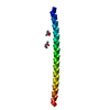
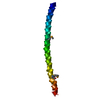
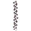
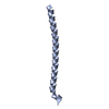
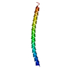
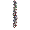
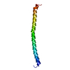
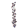
| Title | CRYSTAL STRUCTURE OF THE N-TERMINAL COILED COIL DOMAIN FROM APC | ||||||
 Components Components | ADENOMATOUS POLYPOSIS COLI PROTEIN | ||||||
 Keywords Keywords | STRUCTURAL PROTEIN / APC / COILED COIL / TUMOR SUPPRESSOR | ||||||
| Function / homology |  Function and homology information Function and homology informationAPC truncation mutants are not K63 polyubiquitinated / negative regulation of cell cycle G1/S phase transition / negative regulation of cyclin-dependent protein serine/threonine kinase activity / gamma-catenin binding / regulation of attachment of spindle microtubules to kinetochore / positive regulation of pseudopodium assembly / positive regulation of protein localization to centrosome / bicellular tight junction assembly / pattern specification process / negative regulation of microtubule depolymerization ...APC truncation mutants are not K63 polyubiquitinated / negative regulation of cell cycle G1/S phase transition / negative regulation of cyclin-dependent protein serine/threonine kinase activity / gamma-catenin binding / regulation of attachment of spindle microtubules to kinetochore / positive regulation of pseudopodium assembly / positive regulation of protein localization to centrosome / bicellular tight junction assembly / pattern specification process / negative regulation of microtubule depolymerization / heart valve development / beta-catenin destruction complex / catenin complex / APC truncation mutants have impaired AXIN binding / AXIN missense mutants destabilize the destruction complex / Truncations of AMER1 destabilize the destruction complex / microtubule plus-end binding / protein kinase regulator activity / Beta-catenin phosphorylation cascade / Signaling by GSK3beta mutants / CTNNB1 S33 mutants aren't phosphorylated / CTNNB1 S37 mutants aren't phosphorylated / CTNNB1 S45 mutants aren't phosphorylated / CTNNB1 T41 mutants aren't phosphorylated / Wnt signalosome / regulation of microtubule-based process / cell fate specification / Disassembly of the destruction complex and recruitment of AXIN to the membrane / endocardial cushion morphogenesis / Apoptotic cleavage of cellular proteins / negative regulation of G1/S transition of mitotic cell cycle / mitotic spindle assembly checkpoint signaling / regulation of microtubule-based movement / mitotic cytokinesis / dynein complex binding / lateral plasma membrane / bicellular tight junction / adherens junction / Deactivation of the beta-catenin transactivating complex / negative regulation of canonical Wnt signaling pathway / beta-catenin binding / kinetochore / Degradation of beta-catenin by the destruction complex / ruffle membrane / Wnt signaling pathway / positive regulation of protein catabolic process / insulin receptor signaling pathway / Ovarian tumor domain proteases / nervous system development / cell migration / lamellipodium / positive regulation of cold-induced thermogenesis / protein-containing complex assembly / microtubule binding / microtubule / proteasome-mediated ubiquitin-dependent protein catabolic process / cell adhesion / positive regulation of cell migration / positive regulation of apoptotic process / negative regulation of cell population proliferation / DNA damage response / ubiquitin protein ligase binding / centrosome / protein kinase binding / perinuclear region of cytoplasm / Golgi apparatus / nucleoplasm / nucleus / plasma membrane / cytoplasm / cytosol Similarity search - Function | ||||||
| Biological species |  Homo sapiens (human) Homo sapiens (human) | ||||||
| Method |  X-RAY DIFFRACTION / Resolution: 2.4 Å X-RAY DIFFRACTION / Resolution: 2.4 Å | ||||||
 Authors Authors | Day, C.L. / Alber, T. | ||||||
 Citation Citation | Journal: J.Mol.Biol. / Year: 2000 Title: Crystal structure of the amino-terminal coiled-coil domain of the APC tumor suppressor. Authors: Day, C.L. / Alber, T. | ||||||
| History |
|
- Structure visualization
| Structure viewer | Molecule: MolmilJmol/JSmol |
|---|
- Downloads & links
-Download
| PDBx/mmCIF format | 1deb.cif.gz | 31.8 KB | Display | PDBx/mmCIF format |
|---|---|---|---|---|
| PDB format | pdb1deb.ent.gz | 22.8 KB | Display | PDB format |
| PDBx/mmJSON format | 1deb.json.gz | Tree view | PDBx/mmJSON format | |
| Others |  Other downloads Other downloads |
-Validation report
| Arichive directory | https://data.pdbj.org/pub/pdb/validation_reports/de/1debftp://data.pdbj.org/pub/pdb/validation_reports/de/1deb | HTTPS FTP |
|---|
-Related structure data
| Similar structure data |
|---|


-Links
 PDBj
PDBj


- Assembly
| Deposited unit | 
| ||||||||
|---|---|---|---|---|---|---|---|---|---|
| 1 | 
| ||||||||
| 2 | 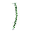
| ||||||||
| 3 | 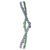
| ||||||||
| 4 |
| ||||||||
| 5 | 
| ||||||||
| Unit cell |
|
-Components
| #1: Protein | Mass: 6109.825 Da / Num. of mol.: 2 / Fragment: N-TERMINAL COILED COIL DOMAIN Source method: isolated from a genetically manipulated source Source: (gene. exp.) Homo sapiens (human) / Plasmid: PAED4 / Production host:  #2: Chemical | ChemComp-SO4 / |   Mass: 96.063 Da / Num. of mol.: 1 / Source method: obtained synthetically / Formula: SO4 Mass: 96.063 Da / Num. of mol.: 1 / Source method: obtained synthetically / Formula: SO4#3: Water | ChemComp-HOH / |  Mass: 18.015 Da / Num. of mol.: 34 / Source method: isolated from a natural source / Formula: H2O Mass: 18.015 Da / Num. of mol.: 34 / Source method: isolated from a natural source / Formula: H2O |
|---|
-Experimental details
-Experiment
| Experiment | Method: X-RAY DIFFRACTION / Number of used crystals: 1 |
|---|
- Sample preparation
| Crystal | Density Matthews: 3.37 Å3/Da / Density % sol: 63.45 % | |||||||||||||||||||||||||
|---|---|---|---|---|---|---|---|---|---|---|---|---|---|---|---|---|---|---|---|---|---|---|---|---|---|---|
| Crystal grow | Temperature: 277 K / Method: vapor diffusion, hanging drop / pH: 4.8 Details: 15% PEG 4000, 0.1M AMMONIUM ACETATE, 0.1M NA ACETATE, pH 4.8, VAPOR DIFFUSION, HANGING DROP | |||||||||||||||||||||||||
| Crystal grow | *PLUS Method: vapor diffusion, sitting drop | |||||||||||||||||||||||||
| Components of the solutions | *PLUS
|
-Data collection
| Diffraction | Mean temperature: 76 K |
|---|---|
| Diffraction source | Source: ROTATING ANODE / Type: RIGAKU RU200 / Wavelength: 1.5418 |
| Detector | Type: RIGAKU RAXIS IIC / Detector: IMAGE PLATE / Date: 1994 |
| Radiation | Protocol: SINGLE WAVELENGTH / Monochromatic (M) / Laue (L): M / Scattering type: x-ray |
| Radiation wavelength | Wavelength: 1.5418 Å / Relative weight: 1 |
| Reflection | Resolution: 2.4→20 Å / Num. all: 18321 / % possible obs: 93.6 % / Redundancy: 2.8 % / Biso Wilson estimate: 34.63 Å2 / Rmerge(I) obs: 0.079 / Net I/σ(I): 5.3 |
| Reflection shell | Resolution: 2.4→2.48 Å / Redundancy: 3.1 % / Rmerge(I) obs: 0.164 / % possible all: 94 |
| Reflection | *PLUS Num. obs: 6743 / % possible obs: 96.4 % / Num. measured all: 18321 / Rmerge(I) obs: 0.078 |
| Reflection shell | *PLUS % possible obs: 96.7 % / Rmerge(I) obs: 0.176 |
- Processing
| Software |
| ||||||||||||||||||||||||||||||||||||||||||||||||||||||||||||
|---|---|---|---|---|---|---|---|---|---|---|---|---|---|---|---|---|---|---|---|---|---|---|---|---|---|---|---|---|---|---|---|---|---|---|---|---|---|---|---|---|---|---|---|---|---|---|---|---|---|---|---|---|---|---|---|---|---|---|---|---|---|
| Refinement | Resolution: 2.4→20 Å / σ(F): 1 / Stereochemistry target values: ENGH & HUBER
| ||||||||||||||||||||||||||||||||||||||||||||||||||||||||||||
| Refinement step | Cycle: LAST / Resolution: 2.4→20 Å
| ||||||||||||||||||||||||||||||||||||||||||||||||||||||||||||
| Refine LS restraints |
| ||||||||||||||||||||||||||||||||||||||||||||||||||||||||||||
| Software | *PLUS Name: CNS / Classification: refinement | ||||||||||||||||||||||||||||||||||||||||||||||||||||||||||||
| Refinement | *PLUS Highest resolution: 2.4 Å / Lowest resolution: 20 Å / σ(F): 1 / Num. reflection obs: 6148 / Rfactor obs: 0.2326 / Rfactor Rfree: 0.2746 | ||||||||||||||||||||||||||||||||||||||||||||||||||||||||||||
| Solvent computation | *PLUS | ||||||||||||||||||||||||||||||||||||||||||||||||||||||||||||
| Displacement parameters | *PLUS |
