 Movie
Movie Controller


+ Open data

- Basic information
| Entry | Database: PDB / ID: 6s8x | ||||||
|---|---|---|---|---|---|---|---|
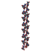
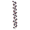
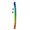
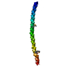
| Title | Crystal structure of the Rab-binding domain of FIP2 | ||||||
 Components Components | Rab11 family-interacting protein 2 | ||||||
 Keywords Keywords | SIGNALING PROTEIN / membrane trafficking / Rab small GTPases / effector protein / Rab-binding domain / endosomal trafficking | ||||||
| Function / homology |  Function and homology information Function and homology informationTRAM-dependent toll-like receptor 4 signaling pathway / regulated exocytosis / insulin secretion involved in cellular response to glucose stimulus / establishment of cell polarity / positive regulation of GTPase activity / phagocytosis / phagocytic cup / cytoplasmic vesicle membrane / positive regulation of protein localization to plasma membrane / small GTPase binding ...TRAM-dependent toll-like receptor 4 signaling pathway / regulated exocytosis / insulin secretion involved in cellular response to glucose stimulus / establishment of cell polarity / positive regulation of GTPase activity / phagocytosis / phagocytic cup / cytoplasmic vesicle membrane / positive regulation of protein localization to plasma membrane / small GTPase binding / recycling endosome membrane / Vasopressin regulates renal water homeostasis via Aquaporins / endosome / protein kinase binding / protein homodimerization activity / nucleoplasm / identical protein binding Similarity search - Function | ||||||
| Biological species |  Homo sapiens (human) Homo sapiens (human) | ||||||
| Method |  X-RAY DIFFRACTION / SYNCHROTRON / AB INITIO PHASING / Resolution: 2.29 Å X-RAY DIFFRACTION / SYNCHROTRON / AB INITIO PHASING / Resolution: 2.29 Å | ||||||
 Authors Authors | Kearney, A.M. / Khan, A.R. | ||||||
| Funding support |  Ireland, 1items Ireland, 1items
| ||||||
 Citation Citation | Journal: Acta Crystallogr.,Sect.F / Year: 2020 Title: Crystal structure of the Rab-binding domain of Rab11 family-interacting protein 2. Authors: Kearney, A.M. / Khan, A.R. | ||||||
| History |
|
- Structure visualization
| Structure viewer | Molecule: MolmilJmol/JSmol |
|---|
- Downloads & links
-Download
| PDBx/mmCIF format | 6s8x.cif.gz | 163.7 KB | Display | PDBx/mmCIF format |
|---|---|---|---|---|
| PDB format | pdb6s8x.ent.gz | 110.9 KB | Display | PDB format |
| PDBx/mmJSON format | 6s8x.json.gz | Tree view | PDBx/mmJSON format | |
| Others |  Other downloads Other downloads |
-Validation report
| Arichive directory | https://data.pdbj.org/pub/pdb/validation_reports/s8/6s8xftp://data.pdbj.org/pub/pdb/validation_reports/s8/6s8x | HTTPS FTP |
|---|
-Related structure data
| Similar structure data |
|---|


-Links
 PDBj
PDBj


- Assembly
| Deposited unit | 
| |||||||||||||||
|---|---|---|---|---|---|---|---|---|---|---|---|---|---|---|---|---|
| 1 | 
| |||||||||||||||
| 2 | 
| |||||||||||||||
| 3 | 
| |||||||||||||||
| 4 | 
| |||||||||||||||
| Unit cell |
| |||||||||||||||
| Components on special symmetry positions |
|
-Components
| #1: Protein | Mass: 9121.314 Da / Num. of mol.: 4 Source method: isolated from a genetically manipulated source Source: (gene. exp.) Homo sapiens (human) / Gene: RAB11FIP2, KIAA0941 / Production host:  #2: Chemical | ChemComp-HEZ / |   Mass: 118.174 Da / Num. of mol.: 1 / Source method: obtained synthetically / Formula: C6H14O2 Mass: 118.174 Da / Num. of mol.: 1 / Source method: obtained synthetically / Formula: C6H14O2#3: Water | ChemComp-HOH / |  Mass: 18.015 Da / Num. of mol.: 62 / Source method: isolated from a natural source / Formula: H2O Mass: 18.015 Da / Num. of mol.: 62 / Source method: isolated from a natural source / Formula: H2OHas ligand of interest | N | |
|---|
-Experimental details
-Experiment
| Experiment | Method: X-RAY DIFFRACTION / Number of used crystals: 1 |
|---|
- Sample preparation
| Crystal | Density Matthews: 3.75 Å3/Da / Density % sol: 67.17 % |
|---|---|
| Crystal grow | Temperature: 290 K / Method: vapor diffusion, sitting drop Details: 0.01M cobat chloride 0.1M NaOAc, pH 4.7 1M 1,6 hexanediol |
-Data collection
| Diffraction | Mean temperature: 100 K / Serial crystal experiment: N |
|---|---|
| Diffraction source | Source: SYNCHROTRON / Site: APS  / Beamline: 24-ID-C / Wavelength: 0.9791 Å / Beamline: 24-ID-C / Wavelength: 0.9791 Å |
| Detector | Type: DECTRIS PILATUS 6M-F / Detector: PIXEL / Date: Aug 12, 2016 |
| Radiation | Protocol: SINGLE WAVELENGTH / Monochromatic (M) / Laue (L): M / Scattering type: x-ray |
| Radiation wavelength | Wavelength: 0.9791 Å / Relative weight: 1 |
| Reflection | Resolution: 2.29→46.16 Å / Num. all: 72768 / Num. obs: 16934 / % possible obs: 99.4 % / Redundancy: 4.3 % / Biso Wilson estimate: 35.97 Å2 / Rmerge(I) obs: 0.111 / Rpim(I) all: 0.059 / Rrim(I) all: 0.126 / Net I/σ(I): 8.8 |
| Reflection shell | Resolution: 2.29→2.37 Å / Redundancy: 4.3 % / Rmerge(I) obs: 0.872 / Mean I/σ(I) obs: 1.7 / Num. unique obs: 1611 / CC1/2: 0.712 / Rpim(I) all: 0.483 / Rrim(I) all: 1.048 / % possible all: 99.3 |
- Processing
| Software |
| |||||||||||||||||||||||||||||||||||||||||||||||||
|---|---|---|---|---|---|---|---|---|---|---|---|---|---|---|---|---|---|---|---|---|---|---|---|---|---|---|---|---|---|---|---|---|---|---|---|---|---|---|---|---|---|---|---|---|---|---|---|---|---|---|
| Refinement | Method to determine structure: AB INITIO PHASING / Resolution: 2.29→43.02 Å / SU ML: 0.2621 / Cross valid method: FREE R-VALUE / σ(F): 1.35 / Phase error: 27.3366
| |||||||||||||||||||||||||||||||||||||||||||||||||
| Solvent computation | Shrinkage radii: 0.9 Å / VDW probe radii: 1.11 Å | |||||||||||||||||||||||||||||||||||||||||||||||||
| Displacement parameters | Biso mean: 45.36 Å2 | |||||||||||||||||||||||||||||||||||||||||||||||||
| Refinement step | Cycle: LAST / Resolution: 2.29→43.02 Å
| |||||||||||||||||||||||||||||||||||||||||||||||||
| Refine LS restraints |
| |||||||||||||||||||||||||||||||||||||||||||||||||
| LS refinement shell |
| |||||||||||||||||||||||||||||||||||||||||||||||||
| Refinement TLS params. | Method: refined / Origin x: 24.7894067103 Å / Origin y: 38.4361092945 Å / Origin z: 66.7265682416 Å
| |||||||||||||||||||||||||||||||||||||||||||||||||
| Refinement TLS group | Selection details: all |
