keratin filament binding / intermediate filament organization / cellular response to muramyl dipeptide / structural constituent of eye lens / intermediate filament cytoskeleton / Striated Muscle Contraction / RHOBTB1 GTPase cycle / intermediate filament / Dengue Virus Genome Translation and Replication / Developmental Lineage of Mammary Gland Myoepithelial Cells ...keratin filament binding / intermediate filament organization / cellular response to muramyl dipeptide / structural constituent of eye lens / intermediate filament cytoskeleton / Striated Muscle Contraction / RHOBTB1 GTPase cycle / intermediate filament / Dengue Virus Genome Translation and Replication / Developmental Lineage of Mammary Gland Myoepithelial Cells / cell leading edge / Dengue Virus-Host Interactions / positive regulation of collagen biosynthetic process / microtubule organizing center / Caspase-mediated cleavage of cytoskeletal proteins / phagocytic vesicle / regulation of mRNA stability / Late endosomal microautophagy / structural constituent of cytoskeleton / nuclear matrix / Chaperone Mediated Autophagy / Aggrephagy / peroxisome / double-stranded RNA binding / cellular response to lipopolysaccharide / Interleukin-4 and Interleukin-13 signaling / scaffold protein binding / molecular adaptor activity / cytoskeleton / protein domain specific binding / axon / focal adhesion / positive regulation of gene expression / extracellular exosome / identical protein binding / plasma membrane / cytosol / cytoplasm Similarity search - Function
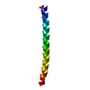
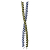
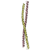
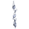
Intermediate filament head, DNA-binding domain / : / Intermediate filament head (DNA binding) region / Intermediate filament protein, conserved site / Intermediate filament protein / Intermediate filament (IF) rod domain signature. / Intermediate filament, rod domain / Intermediate filament (IF) rod domain profile. / Intermediate filament protein / Single alpha-helices involved in coiled-coils or other helix-helix interfaces - #170 ...Intermediate filament head, DNA-binding domain / : / Intermediate filament head (DNA binding) region / Intermediate filament protein, conserved site / Intermediate filament protein / Intermediate filament (IF) rod domain signature. / Intermediate filament, rod domain / Intermediate filament (IF) rod domain profile. / Intermediate filament protein / Single alpha-helices involved in coiled-coils or other helix-helix interfaces - #170 / Single alpha-helices involved in coiled-coils or other helix-helix interfaces / Up-down Bundle / Mainly Alpha Similarity search - Domain/homology
Mass: 18.015 Da / Num. of mol.: 454 / Source method: isolated from a natural source / Formula: H2O
Sequence details
THE RESIDUE NUMBERING USED IN THE LITERATURE FOR VIMENTIN DIFFERS BY +1 FROM THE NUMBERING USED IN ...THE RESIDUE NUMBERING USED IN THE LITERATURE FOR VIMENTIN DIFFERS BY +1 FROM THE NUMBERING USED IN SWISSPROT ENTRY P08670
-
Experimental details
-
Experiment
Experiment
Method: X-RAY DIFFRACTION / Number of used crystals: 1
In the structure databanks used in Yorodumi, some data are registered as the other names, "COVID-19 virus" and "2019-nCoV". Here are the details of the virus and the list of structure data.
Jan 31, 2019. EMDB accession codes are about to change! (news from PDBe EMDB page)
EMDB accession codes are about to change! (news from PDBe EMDB page)
The allocation of 4 digits for EMDB accession codes will soon come to an end. Whilst these codes will remain in use, new EMDB accession codes will include an additional digit and will expand incrementally as the available range of codes is exhausted. The current 4-digit format prefixed with “EMD-” (i.e. EMD-XXXX) will advance to a 5-digit format (i.e. EMD-XXXXX), and so on. It is currently estimated that the 4-digit codes will be depleted around Spring 2019, at which point the 5-digit format will come into force.
The EM Navigator/Yorodumi systems omit the EMD- prefix.
Related info.:Q: What is EMD? / ID/Accession-code notation in Yorodumi/EM Navigator
Yorodumi is a browser for structure data from EMDB, PDB, SASBDB, etc.
This page is also the successor to EM Navigator detail page, and also detail information page/front-end page for Omokage search.
The word "yorodu" (or yorozu) is an old Japanese word meaning "ten thousand". "mi" (miru) is to see.
Related info.:EMDB / PDB / SASBDB / Comparison of 3 databanks / Yorodumi Search / Aug 31, 2016. New EM Navigator & Yorodumi / Yorodumi Papers / Jmol/JSmol / Function and homology information / Changes in new EM Navigator and Yorodumi
 Movie
Movie Controller


 Open data
 Basic information
 Components
 Keywords
 Function and homology information
 HOMO SAPIENS (human)
 X-RAY DIFFRACTION /
  Authors
 Citation
 Structure visualization
 Downloads & links
 Other downloads


 PDBj
PDBj


 Assembly


 Mass: 59.044 Da / Num. of mol.: 1 / Source method: obtained synthetically / Formula: C2H3O2
 Mass: 18.015 Da / Num. of mol.: 454 / Source method: isolated from a natural source / Formula: H2O
 Sample preparation
 / Beamline: BM1A / Wavelength: 0.9
  Processing