 Movie
Movie Controller


+ Open data

- Basic information
| Entry | Database: PDB / ID: 1nyh | ||||||
|---|---|---|---|---|---|---|---|
| Title | Crystal Structure of the Coiled-coil Dimerization Motif of Sir4 | ||||||
 Components Components | Regulatory protein SIR4 | ||||||
 Keywords Keywords | Transcription repressor / coiled-coil / Transcription regulation / Repressor | ||||||
| Function / homology |  Function and homology information Function and homology informationestablishment of protein-containing complex localization to telomere / telomere tethering at nuclear periphery / chromatin silencing complex / silent mating-type cassette heterochromatin formation / positive regulation of heterochromatin formation / subtelomeric heterochromatin formation / nucleosome binding / double-strand break repair via nonhomologous end joining / heterochromatin formation / double-stranded DNA binding ...establishment of protein-containing complex localization to telomere / telomere tethering at nuclear periphery / chromatin silencing complex / silent mating-type cassette heterochromatin formation / positive regulation of heterochromatin formation / subtelomeric heterochromatin formation / nucleosome binding / double-strand break repair via nonhomologous end joining / heterochromatin formation / double-stranded DNA binding / molecular adaptor activity / chromosome, telomeric region Similarity search - Function | ||||||
| Biological species |  | ||||||
| Method |  X-RAY DIFFRACTION / SYNCHROTRON / MAD / Resolution: 3.1 Å X-RAY DIFFRACTION / SYNCHROTRON / MAD / Resolution: 3.1 Å | ||||||
 Authors Authors | Chang, J.F. / Hall, B.E. / Tanny, J.C. / Moazed, D. / Filman, D. / Ellenberger, T. | ||||||
 Citation Citation | Journal: Structure / Year: 2003 Title: Structure of the Coiled-coil Dimerization Motif of Sir4 and Its Interaction With Sir3 Authors: Chang, J.F. / Hall, B.E. / Tanny, J.C. / Moazed, D. / Filman, D. / Ellenberger, T. | ||||||
| History |
|
- Structure visualization
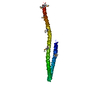
| Structure viewer | Molecule: MolmilJmol/JSmol |
|---|
- Downloads & links
-Download
| PDBx/mmCIF format | 1nyh.cif.gz | 26.9 KB | Display | PDBx/mmCIF format |
|---|---|---|---|---|
| PDB format | pdb1nyh.ent.gz | 17.1 KB | Display | PDB format |
| PDBx/mmJSON format | 1nyh.json.gz | Tree view | PDBx/mmJSON format | |
| Others |  Other downloads Other downloads |
-Validation report
| Arichive directory | https://data.pdbj.org/pub/pdb/validation_reports/ny/1nyhftp://data.pdbj.org/pub/pdb/validation_reports/ny/1nyh | HTTPS FTP |
|---|
-Related structure data
| Similar structure data |
|---|


-Links
 PDBj
PDBj
- Assembly
| Deposited unit | 
| ||||||||
|---|---|---|---|---|---|---|---|---|---|
| 1 | 
| ||||||||
| Unit cell |
| ||||||||
| Details | The biological assembly is a dimer generated from the monomer in the asymmetric unit by the operations: -y, -x, 1/6+z and a translation of +1 along a, b and c axes. |
-Components
| #1: Protein | Mass: 18652.967 Da / Num. of mol.: 1 Fragment: Coiled-coil dimerization domain, residue 1198-1358 Source method: isolated from a genetically manipulated source Source: (gene. exp.) Gene: SIR4 / Plasmid: pGEX6P / Species (production host): Escherichia coli / Production host:  |
|---|
-Experimental details
-Experiment
| Experiment | Method: X-RAY DIFFRACTION / Number of used crystals: 1 |
|---|
- Sample preparation
| Crystal | Density Matthews: 3.49 Å3/Da / Density % sol: 64.78 % | |||||||||||||||||||||||||||||||||||||||||||||||||||||||||||||||
|---|---|---|---|---|---|---|---|---|---|---|---|---|---|---|---|---|---|---|---|---|---|---|---|---|---|---|---|---|---|---|---|---|---|---|---|---|---|---|---|---|---|---|---|---|---|---|---|---|---|---|---|---|---|---|---|---|---|---|---|---|---|---|---|---|
| Crystal grow | Temperature: 295 K / Method: vapor diffusion, hanging drop / pH: 6 Details: PEG 3350, MES, MgCl2, pH 6.0, VAPOR DIFFUSION, HANGING DROP, temperature 295K | |||||||||||||||||||||||||||||||||||||||||||||||||||||||||||||||
| Crystal grow | *PLUS Temperature: 22 ℃ / pH: 7.5 / Method: vapor diffusion, hanging drop | |||||||||||||||||||||||||||||||||||||||||||||||||||||||||||||||
| Components of the solutions | *PLUS
|
-Data collection
| Diffraction | Mean temperature: 110 K | ||||||||||||
|---|---|---|---|---|---|---|---|---|---|---|---|---|---|
| Diffraction source | Source: SYNCHROTRON / Site: NSLS  / Beamline: X12C / Wavelength: 0.97827, 0.978423, 0.95 / Beamline: X12C / Wavelength: 0.97827, 0.978423, 0.95 | ||||||||||||
| Detector | Type: ADSC QUANTUM 4 / Detector: CCD / Date: Jun 21, 2001 / Details: toroidal mirror | ||||||||||||
| Radiation | Monochromator: Channel-Cut Si(111)Monochromator / Protocol: MAD / Monochromatic (M) / Laue (L): M / Scattering type: x-ray | ||||||||||||
| Radiation wavelength |
| ||||||||||||
| Reflection | Resolution: 3.1→47.14 Å / Num. all: 5054 / Num. obs: 4796 / % possible obs: 95 % / Observed criterion σ(F): 1 / Observed criterion σ(I): 1 / Redundancy: 11.2 % / Rmerge(I) obs: 0.066 / Net I/σ(I): 33.3 | ||||||||||||
| Reflection shell | Resolution: 3.1→3.21 Å / Redundancy: 5.6 % / Rmerge(I) obs: 0.303 / Mean I/σ(I) obs: 9.2 / % possible all: 97.2 | ||||||||||||
| Reflection | *PLUS % possible obs: 96.2 % / Num. measured all: 191300 | ||||||||||||
| Reflection shell | *PLUS % possible obs: 97.2 % |
- Processing
| Software |
| ||||||||||||||||||||||||||||||||||||||||||||||||||||||||||||||||||||||||||||||||||||||||||||||||||||||||||||||||||||||||||||||||||
|---|---|---|---|---|---|---|---|---|---|---|---|---|---|---|---|---|---|---|---|---|---|---|---|---|---|---|---|---|---|---|---|---|---|---|---|---|---|---|---|---|---|---|---|---|---|---|---|---|---|---|---|---|---|---|---|---|---|---|---|---|---|---|---|---|---|---|---|---|---|---|---|---|---|---|---|---|---|---|---|---|---|---|---|---|---|---|---|---|---|---|---|---|---|---|---|---|---|---|---|---|---|---|---|---|---|---|---|---|---|---|---|---|---|---|---|---|---|---|---|---|---|---|---|---|---|---|---|---|---|---|---|
| Refinement | Method to determine structure: MAD / Resolution: 3.1→47.14 Å / Cor.coef. Fo:Fc: 0.839 / Cor.coef. Fo:Fc free: 0.833 / SU B: 14.439 / SU ML: 0.254 / Cross valid method: THROUGHOUT / σ(F): 1 / ESU R: 0.425 / ESU R Free: 0.345 Stereochemistry target values: MAXIMUM LIKELIHOOD WITH PHASES
| ||||||||||||||||||||||||||||||||||||||||||||||||||||||||||||||||||||||||||||||||||||||||||||||||||||||||||||||||||||||||||||||||||
| Solvent computation | Ion probe radii: 0.8 Å / Shrinkage radii: 0.8 Å / VDW probe radii: 1.4 Å / Solvent model: BABINET MODEL WITH MASK | ||||||||||||||||||||||||||||||||||||||||||||||||||||||||||||||||||||||||||||||||||||||||||||||||||||||||||||||||||||||||||||||||||
| Displacement parameters | Biso mean: 54.993 Å2
| ||||||||||||||||||||||||||||||||||||||||||||||||||||||||||||||||||||||||||||||||||||||||||||||||||||||||||||||||||||||||||||||||||
| Refinement step | Cycle: LAST / Resolution: 3.1→47.14 Å
| ||||||||||||||||||||||||||||||||||||||||||||||||||||||||||||||||||||||||||||||||||||||||||||||||||||||||||||||||||||||||||||||||||
| Refine LS restraints |
| ||||||||||||||||||||||||||||||||||||||||||||||||||||||||||||||||||||||||||||||||||||||||||||||||||||||||||||||||||||||||||||||||||
| LS refinement shell | Resolution: 3.1→3.184 Å / Total num. of bins used: 20 /
| ||||||||||||||||||||||||||||||||||||||||||||||||||||||||||||||||||||||||||||||||||||||||||||||||||||||||||||||||||||||||||||||||||
| Refinement | *PLUS Rfactor Rfree: 0.288 / Rfactor Rwork: 0.271 | ||||||||||||||||||||||||||||||||||||||||||||||||||||||||||||||||||||||||||||||||||||||||||||||||||||||||||||||||||||||||||||||||||
| Solvent computation | *PLUS | ||||||||||||||||||||||||||||||||||||||||||||||||||||||||||||||||||||||||||||||||||||||||||||||||||||||||||||||||||||||||||||||||||
| Displacement parameters | *PLUS | ||||||||||||||||||||||||||||||||||||||||||||||||||||||||||||||||||||||||||||||||||||||||||||||||||||||||||||||||||||||||||||||||||
| Refine LS restraints | *PLUS
|
