 Movie
Movie Controller

[English] 日本語
 Yorodumi
- PDB-3bat: Crystal structure of the N-terminal region of the scallop myosin ... -
+ Open data

- Basic information
| Entry | Database: PDB / ID: 3bat | ||||||
|---|---|---|---|---|---|---|---|
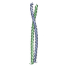
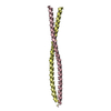
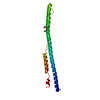
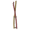
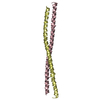
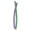
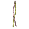
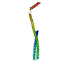
| Title | Crystal structure of the N-terminal region of the scallop myosin rod, monoclinic (P21) form | ||||||
 Components Components | Myosin heavy chain, striated muscle/General control protein GCN4 | ||||||
 Keywords Keywords | CONTRACTILE PROTEIN / Alpha-helical coiled coil / disorder / salt links / Actin-binding / ATP-binding / Calmodulin-binding / Cytoplasm / Motor protein / Muscle protein / Myosin / Nucleotide-binding / Thick filament | ||||||
| Function / homology |  Function and homology information Function and homology informationmyosin filament organization / myofibril assembly / FCERI mediated MAPK activation / protein localization to nuclear periphery / Activation of the AP-1 family of transcription factors / negative regulation of ribosomal protein gene transcription by RNA polymerase II / positive regulation of cellular response to amino acid starvation / response to amino acid starvation / mediator complex binding / myosin filament ...myosin filament organization / myofibril assembly / FCERI mediated MAPK activation / protein localization to nuclear periphery / Activation of the AP-1 family of transcription factors / negative regulation of ribosomal protein gene transcription by RNA polymerase II / positive regulation of cellular response to amino acid starvation / response to amino acid starvation / mediator complex binding / myosin filament / Oxidative Stress Induced Senescence / locomotion / myosin II complex / structural constituent of muscle / microfilament motor activity / amino acid biosynthetic process / TFIID-class transcription factor complex binding / myofibril / positive regulation of RNA polymerase II transcription preinitiation complex assembly / positive regulation of transcription initiation by RNA polymerase II / cellular response to nutrient levels / muscle contraction / cellular response to amino acid starvation / RNA polymerase II transcription regulator complex / actin filament binding / DNA-binding transcription activator activity, RNA polymerase II-specific / transcription regulator complex / sequence-specific DNA binding / RNA polymerase II-specific DNA-binding transcription factor binding / DNA-binding transcription factor activity, RNA polymerase II-specific / calmodulin binding / intracellular signal transduction / RNA polymerase II cis-regulatory region sequence-specific DNA binding / DNA-binding transcription factor activity / chromatin binding / negative regulation of transcription by RNA polymerase II / positive regulation of transcription by RNA polymerase II / ATP binding / identical protein binding / nucleus Similarity search - Function | ||||||
| Biological species |  Argopecten irradians (bay scallop) Argopecten irradians (bay scallop) | ||||||
| Method |  X-RAY DIFFRACTION / SYNCHROTRON / MOLECULAR REPLACEMENT / Resolution: 2.3 Å X-RAY DIFFRACTION / SYNCHROTRON / MOLECULAR REPLACEMENT / Resolution: 2.3 Å | ||||||
 Authors Authors | Brown, J.H. / Cohen, C. | ||||||
 Citation Citation | Journal: J.Mol.Biol. / Year: 2008 Title: An unstable head-rod junction may promote folding into the compact off-state conformation of regulated myosins. Authors: Brown, J.H. / Yang, Y. / Reshetnikova, L. / Gourinath, S. / Suveges, D. / Kardos, J. / Hobor, F. / Reutzel, R. / Nyitray, L. / Cohen, C. #1: Journal: Nature / Year: 2003Title: Visualization of an unstable coiled coil from the scallop myosin rod. Authors: Li, Y. / Brown, J.H. / Reshetnikova, L. / Blazsek, A. / Farkas, L. / Nyitray, L. / Cohen, C. #2: Journal: Proc Natl Acad Sci U S A / Year: 2006Title: Crystal structures of human cardiac beta-myosin II S2-Delta provide insight into the functional role of the S2 subfragment. Authors: Wulf Blankenfeldt / Nicolas H Thomä / John S Wray / Mathias Gautel / Ilme Schlichting /  Abstract: Myosin II is the major component of the muscle thick filament. It consists of two N-terminal S1 subfragments ("heads") connected to a long dimeric coiled-coil rod. The rod is in itself twofold ...Myosin II is the major component of the muscle thick filament. It consists of two N-terminal S1 subfragments ("heads") connected to a long dimeric coiled-coil rod. The rod is in itself twofold symmetric, but in the filament, the two heads point away from the filament surface and are therefore not equivalent. This breaking of symmetry requires the initial section of the rod, subfragment 2 (S2), to be relatively flexible. S2 is an important functional element, involved in various mechanisms by which the activity of smooth and striated muscle is regulated. We have determined crystal structures of the 126 N-terminal residues of S2 from human cardiac beta-myosin II (S2-Delta), of both WT and the disease-associated E924K mutant. S2-Delta is a straight parallel dimeric coiled coil, but the N terminus of one chain is disordered in WT-S2-Delta due to crystal contacts, indicative of unstable local structure. Bulky noncanonical side chains pack into a/d positions of S2-Delta's N terminus, leading to defined local asymmetry and axial stagger, which could induce nonequivalence of the S1 subfragments. Additionally, S2 possesses a conserved charge distribution with three prominent rings of negative potential within S2-Delta, the first of which may provide a binding interface for the "blocked head" of smooth muscle myosin in the OFF state. The observation that many disease-associated mutations affect the second negatively charged ring further suggests that charge interactions play an important role in regulation of cardiac muscle activity through myosin-binding protein C. | ||||||
| History |
| ||||||
| Remark 999 | SEQUENCE THE PEPTIDE CONTAINS A GSHM TETRAPEPTIDE, FOLLOWED BY THE N-TERMINAL 51 RESIDUES OF THE ... SEQUENCE THE PEPTIDE CONTAINS A GSHM TETRAPEPTIDE, FOLLOWED BY THE N-TERMINAL 51 RESIDUES OF THE BAY SCALLOP MYOSIN ROD (RESIDUES 835-885 OF THE BAY SCALLOP MYOSIN HEAVY CHAIN, GI:5612), FOLLOWED BY A GS LINKER, FOLLOWED BY THE LEUCINE ZIPPER OF THE YEAST GCN4 TRANSCRIPTION FACTOR (RESIDUES 250-281 OF GI:171584 DENOTED AS RESIDUES 888-919 IN THE SUBMITTED COORDINATES). |
- Structure visualization
| Structure viewer | Molecule: MolmilJmol/JSmol |
|---|
- Downloads & links
-Download
| PDBx/mmCIF format | 3bat.cif.gz | 80.2 KB | Display | PDBx/mmCIF format |
|---|---|---|---|---|
| PDB format | pdb3bat.ent.gz | 61.9 KB | Display | PDB format |
| PDBx/mmJSON format | 3bat.json.gz | Tree view | PDBx/mmJSON format | |
| Others |  Other downloads Other downloads |
-Validation report
| Arichive directory | https://data.pdbj.org/pub/pdb/validation_reports/ba/3batftp://data.pdbj.org/pub/pdb/validation_reports/ba/3bat | HTTPS FTP |
|---|
-Related structure data
| Related structure data |  3basC 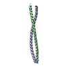 1nknS C: citing same article ( S: Starting model for refinement |
|---|---|
| Similar structure data |


-Links
 PDBj
PDBj


- Assembly
| Deposited unit | 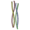
| ||||||||
|---|---|---|---|---|---|---|---|---|---|
| 1 | 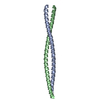
| ||||||||
| 2 | 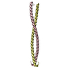
| ||||||||
| Unit cell |
|
-Components
| #1: Protein | Mass: 10559.174 Da / Num. of mol.: 4 Fragment: Bay Scallop Myosin (Residues 835-885)/Yeast GCN4 Transcription Factor (Residues 250-281) Source method: isolated from a genetically manipulated source Source: (gene. exp.) Argopecten irradians (bay scallop), (gene. exp.) Genus: Argopecten, Saccharomyces / Species: , / Strain: , / Tissue: Adductor muscle/- / Gene: -/GCN4, AAS3, ARG9, YEL009C / Plasmid: pET15b / Production host:  #2: Water | ChemComp-HOH / |  Mass: 18.015 Da / Num. of mol.: 99 / Source method: isolated from a natural source / Formula: H2O Mass: 18.015 Da / Num. of mol.: 99 / Source method: isolated from a natural source / Formula: H2O |
|---|
-Experimental details
-Experiment
| Experiment | Method: X-RAY DIFFRACTION / Number of used crystals: 1 |
|---|
- Sample preparation
| Crystal | Density Matthews: 2.25 Å3/Da / Density % sol: 45.45 % |
|---|---|
| Crystal grow | Temperature: 277 K / Method: vapor diffusion, hanging drop / pH: 6.2 Details: 2 microliters of protein solution (4 mg/ml protein in 30 mM MOPS buffer pH 7.2, 40 mM NaCl, 2 mM NaN3) mixed with 2 microliters of (25% PEG 3350, 50 mM NH4I) and equilibrated against 1 ml of ...Details: 2 microliters of protein solution (4 mg/ml protein in 30 mM MOPS buffer pH 7.2, 40 mM NaCl, 2 mM NaN3) mixed with 2 microliters of (25% PEG 3350, 50 mM NH4I) and equilibrated against 1 ml of (17.5% PEG 3350, 35 mM NH4I, 28 mM NaCl, 2 mM NaN3, 20 mM MOPS pH 6.2). Harvested crystals were cryoprotected in 25.5% PEG 3350 and 15% glycerol, VAPOR DIFFUSION, HANGING DROP, temperature 277K |
-Data collection
| Diffraction | Mean temperature: 100 K | |||||||||||||||||||||||||||||||||||||||||||||||||||||||
|---|---|---|---|---|---|---|---|---|---|---|---|---|---|---|---|---|---|---|---|---|---|---|---|---|---|---|---|---|---|---|---|---|---|---|---|---|---|---|---|---|---|---|---|---|---|---|---|---|---|---|---|---|---|---|---|---|
| Diffraction source | Source: SYNCHROTRON / Site: NSLS  / Beamline: X26C / Wavelength: 0.9791 Å / Beamline: X26C / Wavelength: 0.9791 Å | |||||||||||||||||||||||||||||||||||||||||||||||||||||||
| Detector | Type: ADSC QUANTUM 4 / Detector: CCD Details: channel-cut Si(111) crystal monochromator followed by a doubly focusing toroidal mirror | |||||||||||||||||||||||||||||||||||||||||||||||||||||||
| Radiation | Monochromator: Si(111) crystal / Protocol: SINGLE WAVELENGTH / Monochromatic (M) / Laue (L): M / Scattering type: x-ray | |||||||||||||||||||||||||||||||||||||||||||||||||||||||
| Radiation wavelength | Wavelength: 0.9791 Å / Relative weight: 1 | |||||||||||||||||||||||||||||||||||||||||||||||||||||||
| Reflection | Resolution: 1.95→30 Å / Num. obs: 27340 / % possible obs: 98.5 % / Rmerge(I) obs: 0.052 / Χ2: 1.085 / Net I/σ(I): 13.8 | |||||||||||||||||||||||||||||||||||||||||||||||||||||||
| Reflection shell |
|
- Processing
| Software |
| ||||||||||||||||||||||||
|---|---|---|---|---|---|---|---|---|---|---|---|---|---|---|---|---|---|---|---|---|---|---|---|---|---|
| Refinement | Method to determine structure: MOLECULAR REPLACEMENT Starting model: PDB entry 1NKN Resolution: 2.3→30 Å / Cross valid method: THROUGHOUT / σ(F): 0
| ||||||||||||||||||||||||
| Solvent computation | Bsol: 38.499 Å2 | ||||||||||||||||||||||||
| Displacement parameters | Biso mean: 53.827 Å2
| ||||||||||||||||||||||||
| Refine analyze |
| ||||||||||||||||||||||||
| Refinement step | Cycle: LAST / Resolution: 2.3→30 Å
| ||||||||||||||||||||||||
| Refine LS restraints |
| ||||||||||||||||||||||||
| LS refinement shell | Resolution: 2.3→2.44 Å
| ||||||||||||||||||||||||
| Xplor file |
|
