 Movie
Movie Controller


+ Open data

- Basic information
| Entry | Database: PDB / ID: 5lxo | ||||||
|---|---|---|---|---|---|---|---|
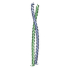
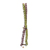
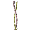
| Title | Coiled-coil protein | ||||||
 Components Components | Transforming acidic coiled-coil-containing protein 3 | ||||||
 Keywords Keywords | STRUCTURAL PROTEIN / TACC3 / coiled-coil / helix / fgfr fusion / fgfr / growth factors / cancer / fusion / parallel helix | ||||||
| Function / homology |  Function and homology information Function and homology informationmicrotubule cytoskeleton organization involved in mitosis / metaphase/anaphase transition of mitotic cell cycle / nuclear migration / centriolar satellite / mitotic spindle organization / regulation of mitotic spindle organization / NOTCH3 Activation and Transmission of Signal to the Nucleus / cerebral cortex development / Negative regulation of NOTCH4 signaling / microtubule cytoskeleton organization ...microtubule cytoskeleton organization involved in mitosis / metaphase/anaphase transition of mitotic cell cycle / nuclear migration / centriolar satellite / mitotic spindle organization / regulation of mitotic spindle organization / NOTCH3 Activation and Transmission of Signal to the Nucleus / cerebral cortex development / Negative regulation of NOTCH4 signaling / microtubule cytoskeleton organization / spindle pole / mitotic spindle / ciliary basal body / cell division / Golgi apparatus / cytosol / cytoplasm Similarity search - Function | ||||||
| Biological species |  Homo sapiens (human) Homo sapiens (human) | ||||||
| Method |  X-RAY DIFFRACTION / SYNCHROTRON / MOLECULAR REPLACEMENT / Resolution: 2.179 Å X-RAY DIFFRACTION / SYNCHROTRON / MOLECULAR REPLACEMENT / Resolution: 2.179 Å | ||||||
 Authors Authors | Thiyagarajan, N. / Bunney, T.D. / Katan, M. | ||||||
| Funding support |  United Kingdom, 1items United Kingdom, 1items
| ||||||
 Citation Citation | Journal: To Be Published Title: Coiled-coil protein Authors: Thiyagarajan, N. / Bunney, T.D. / Katan, M. | ||||||
| History |
|
- Structure visualization
| Structure viewer | Molecule: MolmilJmol/JSmol |
|---|
- Downloads & links
-Download
| PDBx/mmCIF format | 5lxo.cif.gz | 147.9 KB | Display | PDBx/mmCIF format |
|---|---|---|---|---|
| PDB format | pdb5lxo.ent.gz | 119.5 KB | Display | PDB format |
| PDBx/mmJSON format | 5lxo.json.gz | Tree view | PDBx/mmJSON format | |
| Others |  Other downloads Other downloads |
-Validation report
| Arichive directory | https://data.pdbj.org/pub/pdb/validation_reports/lx/5lxoftp://data.pdbj.org/pub/pdb/validation_reports/lx/5lxo | HTTPS FTP |
|---|
-Related structure data
| Related structure data |  5lxnSC S: Starting model for refinement C: citing same article ( |
|---|---|
| Similar structure data |


-Links
 PDBj
PDBj
- Assembly
| Deposited unit | 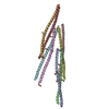
| ||||||||
|---|---|---|---|---|---|---|---|---|---|
| 1 | 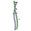
| ||||||||
| 2 | 
| ||||||||
| 3 | 
| ||||||||
| 4 | 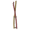
| ||||||||
| Unit cell |
|
-Components
-Protein , 1 types, 8 molecules ABCDEFGH
| #1: Protein | Mass: 9448.420 Da / Num. of mol.: 8 Source method: isolated from a genetically manipulated source Details: Coiled-coil 2 domain / Source: (gene. exp.) Homo sapiens (human) / Gene: TACC3, ERIC1 / Plasmid: pJ821 / Details (production host): Rhamnose induced / Production host:  |
|---|
-Non-polymers , 5 types, 392 molecules 


| #2: Chemical | ChemComp-SO4 /  Mass: 96.063 Da / Num. of mol.: 1 / Source method: obtained synthetically / Formula: SO4 Mass: 96.063 Da / Num. of mol.: 1 / Source method: obtained synthetically / Formula: SO4 | ||||||
|---|---|---|---|---|---|---|---|
| #3: Chemical |  Mass: 150.173 Da / Num. of mol.: 2 / Source method: obtained synthetically / Formula: C6H14O4 Mass: 150.173 Da / Num. of mol.: 2 / Source method: obtained synthetically / Formula: C6H14O4#4: Chemical | ChemComp-ACT / |  Mass: 59.044 Da / Num. of mol.: 1 / Source method: obtained synthetically / Formula: C2H3O2 Mass: 59.044 Da / Num. of mol.: 1 / Source method: obtained synthetically / Formula: C2H3O2#5: Chemical | ChemComp-CL / |  Mass: 35.453 Da / Num. of mol.: 1 / Source method: obtained synthetically / Formula: Cl Mass: 35.453 Da / Num. of mol.: 1 / Source method: obtained synthetically / Formula: Cl#6: Water | ChemComp-HOH / | Mass: 18.015 Da / Num. of mol.: 387 / Source method: isolated from a natural source / Formula: H2O |
-Details
| Has protein modification | Y |
|---|
-Experimental details
-Experiment
| Experiment | Method: X-RAY DIFFRACTION / Number of used crystals: 1 |
|---|
- Sample preparation
| Crystal | Density Matthews: 2.54 Å3/Da / Density % sol: 51.6 % |
|---|---|
| Crystal grow | Temperature: 289.15 K / Method: vapor diffusion, sitting drop / pH: 8.5 Details: 35 % PEG 400, 0.05 M Sodium sulphate, 0.05 M Lithium sulphate, 0.05 M Tris pH 8.5 |
-Data collection
| Diffraction | Mean temperature: 100 K |
|---|---|
| Diffraction source | Source: SYNCHROTRON / Site: Diamond / Beamline: I03 / Wavelength: 0.9681 Å |
| Detector | Type: DECTRIS PILATUS3 6M / Detector: PIXEL / Date: Mar 7, 2015 / Details: Mirrors |
| Radiation | Monochromator: Si / Protocol: SINGLE WAVELENGTH / Monochromatic (M) / Laue (L): M / Scattering type: x-ray |
| Radiation wavelength | Wavelength: 0.9681 Å / Relative weight: 1 |
| Reflection | Resolution: 2.179→28.964 Å / Num. obs: 37534 / % possible obs: 95.5 % / Redundancy: 13.1 % / Biso Wilson estimate: 19.5 Å2 / CC1/2: 0.999 / Rmerge(I) obs: 0.094 / Net I/σ(I): 21 |
| Reflection shell | Resolution: 2.18→2.24 Å / Redundancy: 7.3 % / Rmerge(I) obs: 0.667 / Mean I/σ(I) obs: 2.3 / CC1/2: 0.838 / % possible all: 67.8 |
- Processing
| Software |
| ||||||||||||||||||||||||||||||||||||||||||||||||||||||||||||||||||||||||||||||||||||||||||||||||||
|---|---|---|---|---|---|---|---|---|---|---|---|---|---|---|---|---|---|---|---|---|---|---|---|---|---|---|---|---|---|---|---|---|---|---|---|---|---|---|---|---|---|---|---|---|---|---|---|---|---|---|---|---|---|---|---|---|---|---|---|---|---|---|---|---|---|---|---|---|---|---|---|---|---|---|---|---|---|---|---|---|---|---|---|---|---|---|---|---|---|---|---|---|---|---|---|---|---|---|---|
| Refinement | Method to determine structure: MOLECULAR REPLACEMENT Starting model: 5LXN Resolution: 2.179→28.964 Å / Cross valid method: FREE R-VALUE / σ(F): 1.35 / Phase error: 36.38
| ||||||||||||||||||||||||||||||||||||||||||||||||||||||||||||||||||||||||||||||||||||||||||||||||||
| Solvent computation | Shrinkage radii: 0.9 Å / VDW probe radii: 1.11 Å | ||||||||||||||||||||||||||||||||||||||||||||||||||||||||||||||||||||||||||||||||||||||||||||||||||
| Displacement parameters | Biso mean: 42.4 Å2 | ||||||||||||||||||||||||||||||||||||||||||||||||||||||||||||||||||||||||||||||||||||||||||||||||||
| Refinement step | Cycle: LAST / Resolution: 2.179→28.964 Å
| ||||||||||||||||||||||||||||||||||||||||||||||||||||||||||||||||||||||||||||||||||||||||||||||||||
| Refine LS restraints |
| ||||||||||||||||||||||||||||||||||||||||||||||||||||||||||||||||||||||||||||||||||||||||||||||||||
| LS refinement shell |
|
