 Movie
Movie Controller


+ Open data

- Basic information
| Entry | Database: PDB / ID: 1ic2 | ||||||
|---|---|---|---|---|---|---|---|
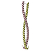
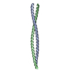
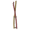
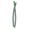
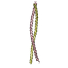
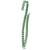
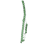
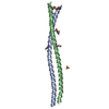
| Title | DECIPHERING THE DESIGN OF THE TROPOMYOSIN MOLECULE | ||||||
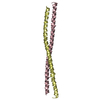
 Components Components | TROPOMYOSIN ALPHA CHAIN, SKELETAL MUSCLE | ||||||
 Keywords Keywords | CONTRACTILE PROTEIN / alpha-helical coiled coil / alanine / symmetry / axial stagger / bend | ||||||
| Function / homology |  Function and homology information Function and homology informationSmooth Muscle Contraction / Striated Muscle Contraction / cytoskeletal protein binding / cardiac muscle contraction / actin filament organization / actin filament / actin filament binding / actin cytoskeleton / protein heterodimerization activity / protein homodimerization activity ...Smooth Muscle Contraction / Striated Muscle Contraction / cytoskeletal protein binding / cardiac muscle contraction / actin filament organization / actin filament / actin filament binding / actin cytoskeleton / protein heterodimerization activity / protein homodimerization activity / identical protein binding / plasma membrane Similarity search - Function | ||||||
| Biological species |  | ||||||
| Method |  X-RAY DIFFRACTION / SYNCHROTRON / MIR / Resolution: 2 Å X-RAY DIFFRACTION / SYNCHROTRON / MIR / Resolution: 2 Å | ||||||
 Authors Authors | Brown, J.H. / Kim, K.-H. / Jun, G. / Greenfield, N.J. / Dominguez, R. / Volkmann, N. / Hitchcock-DeGregori, S.E. / Cohen, C. | ||||||
 Citation Citation | Journal: Proc.Natl.Acad.Sci.USA / Year: 2001 Title: Deciphering the design of the tropomyosin molecule Authors: Brown, J.H. / Kim, K.-H. / Jun, G. / Greenfield, N.J. / Dominguez, R. / Volkmann, N. / Hitchcock-DeGregori, S.E. / Cohen, C. #1: Journal: J.Mol.Biol. / Year: 1969Title: Tropomyosin: Crystal Structure, Polymorphism and Molecular Interactions Authors: Caspar, D.L. / Cohen, C. / Longley, W. #2: Journal: J.Mol.Biol. / Year: 1986Title: Tropomyosin Crystal Structure and Muscle Regulation Authors: Phillips Jr., G.N. / Fillers, J.P. / Cohen, C. #3: Journal: Biochemistry / Year: 1998Title: The Structure of the N-terminus of Striated Muscle alpha-Tropomyosin in a Chimeric Peptide: Nuclear Magnetic Resonance Structure and Circular Dichroism Studies Authors: Greenfield, N.J. / Montelione, G.T. / Farid, R.S. / Hitchcock-DeGregori, S.E. #4: Journal: Proteins / Year: 2000Title: Crystal Structure of Tropomyosin at 7 Angstroms Resolution Authors: Whitby, F.G. / Phillips Jr., G.N. | ||||||
| History |
|
- Structure visualization
| Structure viewer | Molecule: MolmilJmol/JSmol |
|---|
- Downloads & links
-Download
| PDBx/mmCIF format | 1ic2.cif.gz | 74.7 KB | Display | PDBx/mmCIF format |
|---|---|---|---|---|
| PDB format | pdb1ic2.ent.gz | 58.5 KB | Display | PDB format |
| PDBx/mmJSON format | 1ic2.json.gz | Tree view | PDBx/mmJSON format | |
| Others |  Other downloads Other downloads |
-Validation report
| Arichive directory | https://data.pdbj.org/pub/pdb/validation_reports/ic/1ic2ftp://data.pdbj.org/pub/pdb/validation_reports/ic/1ic2 | HTTPS FTP |
|---|
-Related structure data
| Similar structure data |
|---|


-Links
 PDBj
PDBj- Assembly
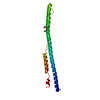
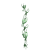
| Deposited unit | 
| ||||||||
|---|---|---|---|---|---|---|---|---|---|
| 1 | 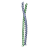
| ||||||||
| 2 | 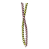
| ||||||||
| Unit cell |
|
-Components
| #1: Protein | Mass: 9273.543 Da / Num. of mol.: 4 / Mutation: A81S Source method: isolated from a genetically manipulated source Source: (gene. exp.)  #2: Water | ChemComp-HOH / |  Mass: 18.015 Da / Num. of mol.: 178 / Source method: isolated from a natural source / Formula: H2O Mass: 18.015 Da / Num. of mol.: 178 / Source method: isolated from a natural source / Formula: H2O |
|---|
-Experimental details
-Experiment
| Experiment | Method: X-RAY DIFFRACTION / Number of used crystals: 5 |
|---|
- Sample preparation
| Crystal | Density Matthews: 2.73 Å3/Da / Density % sol: 54.89 % | ||||||||||||||||||||||||||||||||||||||||||
|---|---|---|---|---|---|---|---|---|---|---|---|---|---|---|---|---|---|---|---|---|---|---|---|---|---|---|---|---|---|---|---|---|---|---|---|---|---|---|---|---|---|---|---|
| Crystal grow | Temperature: 277 K / Method: vapor diffusion / pH: 7.6 Details: PEG 200, isopropanol, magnesium chloride, ammonium acetate, TRIS, pH 7.6, VAPOR DIFFUSION, temperature 277K | ||||||||||||||||||||||||||||||||||||||||||
| Crystal | *PLUS Density % sol: 56 % | ||||||||||||||||||||||||||||||||||||||||||
| Crystal grow | *PLUS Details: used macroseeding | ||||||||||||||||||||||||||||||||||||||||||
| Components of the solutions | *PLUS
|
-Data collection
| Diffraction |
| ||||||||||||||||||||
|---|---|---|---|---|---|---|---|---|---|---|---|---|---|---|---|---|---|---|---|---|---|
| Diffraction source |
| ||||||||||||||||||||
| Detector |
| ||||||||||||||||||||
| Radiation | Protocol: SINGLE WAVELENGTH / Monochromatic (M) / Laue (L): M / Scattering type: x-ray | ||||||||||||||||||||
| Radiation wavelength |
| ||||||||||||||||||||
| Reflection | Resolution: 2→50 Å / Num. all: 26797 / Num. obs: 26797 / % possible obs: 99.9 % / Observed criterion σ(F): 0 / Observed criterion σ(I): 0 / Redundancy: 10 % / Biso Wilson estimate: 31.1 Å2 / Rmerge(I) obs: 0.087 / Net I/σ(I): 15.4 | ||||||||||||||||||||
| Reflection shell | Resolution: 2→2.1 Å / Redundancy: 7.6 % / Rmerge(I) obs: 0.139 / Mean I/σ(I) obs: 10 / % possible all: 99.5 | ||||||||||||||||||||
| Reflection | *PLUS |

- Processing
| Software |
| ||||||||||||||||||||||||||||||||||||
|---|---|---|---|---|---|---|---|---|---|---|---|---|---|---|---|---|---|---|---|---|---|---|---|---|---|---|---|---|---|---|---|---|---|---|---|---|---|
| Refinement | Method to determine structure: MIR / Resolution: 2→50 Å / Isotropic thermal model: anisotropic / Cross valid method: THROUGHOUT / σ(F): 0 / σ(I): 0
| ||||||||||||||||||||||||||||||||||||
| Displacement parameters | Biso mean: 43.8603 Å2
| ||||||||||||||||||||||||||||||||||||
| Refine analyze |
| ||||||||||||||||||||||||||||||||||||
| Refinement step | Cycle: LAST / Resolution: 2→50 Å
| ||||||||||||||||||||||||||||||||||||
| Refine LS restraints |
| ||||||||||||||||||||||||||||||||||||
| LS refinement shell |
| ||||||||||||||||||||||||||||||||||||
| Software | *PLUS Name: CNS / Version: 0.9 / Classification: refinement | ||||||||||||||||||||||||||||||||||||
| Refinement | *PLUS Highest resolution: 2 Å / Lowest resolution: 50 Å / σ(F): 0 / % reflection Rfree: 5 % / Rfactor obs: 0.249 | ||||||||||||||||||||||||||||||||||||
| Solvent computation | *PLUS | ||||||||||||||||||||||||||||||||||||
| Displacement parameters | *PLUS | ||||||||||||||||||||||||||||||||||||
| Refine LS restraints | *PLUS
|
