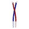
structural constituent of nuclear lamina / negative regulation of mesenchymal cell proliferation / protein localization to nuclear envelope / establishment or maintenance of microtubule cytoskeleton polarity / Breakdown of the nuclear lamina / ventricular cardiac muscle cell development / Depolymerization of the Nuclear Lamina / Nuclear Envelope Breakdown / nuclear pore localization / DNA double-strand break attachment to nuclear envelope ...structural constituent of nuclear lamina / negative regulation of mesenchymal cell proliferation / protein localization to nuclear envelope / establishment or maintenance of microtubule cytoskeleton polarity / Breakdown of the nuclear lamina / ventricular cardiac muscle cell development / Depolymerization of the Nuclear Lamina / Nuclear Envelope Breakdown / nuclear pore localization / DNA double-strand break attachment to nuclear envelope / lamin filament / nuclear envelope organization / XBP1(S) activates chaperone genes / nuclear lamina / Initiation of Nuclear Envelope (NE) Reformation / regulation of protein localization to nucleus / regulation of telomere maintenance / intermediate filament / negative regulation of cardiac muscle hypertrophy in response to stress / muscle organ development / nuclear migration / Deregulated CDK5 triggers multiple neurodegenerative pathways in Alzheimer's disease models / negative regulation of release of cytochrome c from mitochondria / protein localization to nucleus / regulation of cell migration / Meiotic synapsis / negative regulation of extrinsic apoptotic signaling pathway / regulation of protein stability / structural constituent of cytoskeleton / double-strand break repair via nonhomologous end joining / nuclear matrix / protein import into nucleus / cellular senescence / Signaling by BRAF and RAF1 fusions / intracellular protein localization / nuclear envelope / heterochromatin formation / site of double-strand break / nuclear membrane / cellular response to hypoxia / nuclear speck / negative regulation of cell population proliferation / positive regulation of gene expression / perinuclear region of cytoplasm / structural molecule activity / nucleoplasm / identical protein binding / nucleus / cytosol Similarity search - Function
Lamin tail domain superfamily / Lamin tail domain / Lamin Tail Domain / Lamin-tail (LTD) domain profile. / Intermediate filament protein, conserved site / Intermediate filament protein / Intermediate filament (IF) rod domain signature. / Intermediate filament, rod domain / Intermediate filament (IF) rod domain profile. / Intermediate filament protein ...Lamin tail domain superfamily / Lamin tail domain / Lamin Tail Domain / Lamin-tail (LTD) domain profile. / Intermediate filament protein, conserved site / Intermediate filament protein / Intermediate filament (IF) rod domain signature. / Intermediate filament, rod domain / Intermediate filament (IF) rod domain profile. / Intermediate filament protein / Single alpha-helices involved in coiled-coils or other helix-helix interfaces - #170 / Single alpha-helices involved in coiled-coils or other helix-helix interfaces / Up-down Bundle / Mainly Alpha Similarity search - Domain/homology
In the structure databanks used in Yorodumi, some data are registered as the other names, "COVID-19 virus" and "2019-nCoV". Here are the details of the virus and the list of structure data.
Jan 31, 2019. EMDB accession codes are about to change! (news from PDBe EMDB page)
EMDB accession codes are about to change! (news from PDBe EMDB page)
The allocation of 4 digits for EMDB accession codes will soon come to an end. Whilst these codes will remain in use, new EMDB accession codes will include an additional digit and will expand incrementally as the available range of codes is exhausted. The current 4-digit format prefixed with “EMD-” (i.e. EMD-XXXX) will advance to a 5-digit format (i.e. EMD-XXXXX), and so on. It is currently estimated that the 4-digit codes will be depleted around Spring 2019, at which point the 5-digit format will come into force.
The EM Navigator/Yorodumi systems omit the EMD- prefix.
Related info.:Q: What is EMD? / ID/Accession-code notation in Yorodumi/EM Navigator
Yorodumi is a browser for structure data from EMDB, PDB, SASBDB, etc.
This page is also the successor to EM Navigator detail page, and also detail information page/front-end page for Omokage search.
The word "yorodu" (or yorozu) is an old Japanese word meaning "ten thousand". "mi" (miru) is to see.
Related info.:EMDB / PDB / SASBDB / Comparison of 3 databanks / Yorodumi Search / Aug 31, 2016. New EM Navigator & Yorodumi / Yorodumi Papers / Jmol/JSmol / Function and homology information / Changes in new EM Navigator and Yorodumi
 Movie
Movie Controller


 Open data
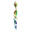
 Basic information
 Components
 Keywords
 Function and homology information
 Homo sapiens (human)
 X-RAY DIFFRACTION /
  Authors
 Citation
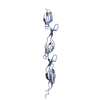
 Structure visualization
 Downloads & links
 Other downloads


 PDBj
PDBj


 Assembly
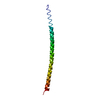

 Sample preparation
 / Beamline: ID23-1 / Wavelength: 0.9769 Å
 Processing