 Movie
Movie Controller

[English] 日本語
 Yorodumi
- PDB-2z5h: Crystal structure of the head-to-tail junction of tropomyosin com... -
+ Open data

- Basic information
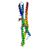
| Entry | Database: PDB / ID: 2z5h | ||||||
|---|---|---|---|---|---|---|---|
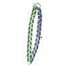
| Title | Crystal structure of the head-to-tail junction of tropomyosin complexed with a fragment of TnT | ||||||
 Components Components |
| ||||||
 Keywords Keywords | CONTRACTILE PROTEIN / actin / troponin / tropomyosin / cytoskeleton / cardiomyopathy / four-helix bundle | ||||||
| Function / homology |  Function and homology information Function and homology informationtroponin C binding / troponin complex / FCERI mediated MAPK activation / protein localization to nuclear periphery / Activation of the AP-1 family of transcription factors / regulation of muscle contraction / negative regulation of ribosomal protein gene transcription by RNA polymerase II / positive regulation of cellular response to amino acid starvation / response to amino acid starvation / mediator complex binding ...troponin C binding / troponin complex / FCERI mediated MAPK activation / protein localization to nuclear periphery / Activation of the AP-1 family of transcription factors / regulation of muscle contraction / negative regulation of ribosomal protein gene transcription by RNA polymerase II / positive regulation of cellular response to amino acid starvation / response to amino acid starvation / mediator complex binding / Oxidative Stress Induced Senescence / sarcomere organization / tropomyosin binding / troponin I binding / amino acid biosynthetic process / TFIID-class transcription factor complex binding / positive regulation of RNA polymerase II transcription preinitiation complex assembly / positive regulation of transcription initiation by RNA polymerase II / skeletal muscle contraction / cellular response to nutrient levels / cellular response to amino acid starvation / actin filament / RNA polymerase II transcription regulator complex / actin filament binding / actin cytoskeleton / DNA-binding transcription activator activity, RNA polymerase II-specific / transcription regulator complex / sequence-specific DNA binding / RNA polymerase II-specific DNA-binding transcription factor binding / DNA-binding transcription factor activity, RNA polymerase II-specific / intracellular signal transduction / RNA polymerase II cis-regulatory region sequence-specific DNA binding / DNA-binding transcription factor activity / protein heterodimerization activity / chromatin binding / negative regulation of transcription by RNA polymerase II / protein homodimerization activity / positive regulation of transcription by RNA polymerase II / identical protein binding / nucleus Similarity search - Function | ||||||
| Biological species |    | ||||||
| Method |  X-RAY DIFFRACTION / SYNCHROTRON / MOLECULAR REPLACEMENT / Resolution: 2.89 Å X-RAY DIFFRACTION / SYNCHROTRON / MOLECULAR REPLACEMENT / Resolution: 2.89 Å | ||||||
 Authors Authors | Murakami, K. / Nozawa, K. / Tomii, K. / Kudou, N. / Igarashi, N. / Shirakihara, Y. / Wakatsuki, S. / Stewart, M. / Yasunaga, T. / Wakabayashi, T. | ||||||
 Citation Citation | Journal: Proc.Natl.Acad.Sci.USA / Year: 2008 Title: Structural basis for tropomyosin overlap in thin (actin) filaments and the generation of a molecular swivel by troponin-T Authors: Murakami, K. / Stewart, M. / Nozawa, K. / Tomii, K. / Kudou, N. / Igarashi, N. / Shirakihara, Y. / Wakatsuki, S. / Yasunaga, T. / Wakabayashi, T. | ||||||
| History |
|
- Structure visualization
| Structure viewer | Molecule: MolmilJmol/JSmol |
|---|
- Downloads & links
-Download
| PDBx/mmCIF format | 2z5h.cif.gz | 106.2 KB | Display | PDBx/mmCIF format |
|---|---|---|---|---|
| PDB format | pdb2z5h.ent.gz | 85.2 KB | Display | PDB format |
| PDBx/mmJSON format | 2z5h.json.gz | Tree view | PDBx/mmJSON format | |
| Others |  Other downloads Other downloads |
-Validation report
| Arichive directory | https://data.pdbj.org/pub/pdb/validation_reports/z5/2z5hftp://data.pdbj.org/pub/pdb/validation_reports/z5/2z5h | HTTPS FTP |
|---|
-Related structure data


-Links
 PDBj
PDBj


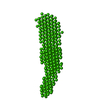
- Assembly
| Deposited unit | 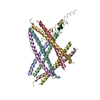
| ||||||||
|---|---|---|---|---|---|---|---|---|---|
| 1 | 
| ||||||||
| 2 | 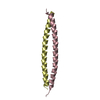
| ||||||||
| 3 | 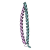
| ||||||||
| 4 | 
| ||||||||
| Unit cell |
|
-Components
| #1: Protein | Mass: 6056.825 Da / Num. of mol.: 8 Fragment: C terminal domain of GCN4 and Tropomyosin alpha-1 chain Source method: isolated from a genetically manipulated source Source: (gene. exp.) Genus: Saccharomyces, Oryctolagus / Species: , / Strain: , / Plasmid: pET3a / Production host:  #2: Protein/peptide | | Mass: 4537.393 Da / Num. of mol.: 1 Fragment: N terminal domain of Tropomyosin alpha-1 chain and C terminal domain of GCN4 Source method: isolated from a genetically manipulated source Source: (gene. exp.) Genus: Oryctolagus, Saccharomyces / Species: , / Strain: , / Plasmid: pET3a / Production host: #3: Protein | | Mass: 6665.503 Da / Num. of mol.: 1 / Fragment: Residues 58-112 Source method: isolated from a genetically manipulated source Source: (gene. exp.) #4: Water | ChemComp-HOH / |  Mass: 18.015 Da / Num. of mol.: 55 / Source method: isolated from a natural source / Formula: H2O Mass: 18.015 Da / Num. of mol.: 55 / Source method: isolated from a natural source / Formula: H2O |
|---|
-Experimental details
-Experiment
| Experiment | Method: X-RAY DIFFRACTION / Number of used crystals: 1 |
|---|
- Sample preparation
| Crystal | Density Matthews: 4.71 Å3/Da / Density % sol: 73.88 % |
|---|---|
| Crystal grow | Temperature: 293 K / Method: vapor diffusion, hanging drop / pH: 6.5 Details: pH 6.5, VAPOR DIFFUSION, HANGING DROP, temperature 293K |
-Data collection
| Diffraction | Mean temperature: 93 K |
|---|---|
| Diffraction source | Source: SYNCHROTRON / Site: Photon Factory  / Beamline: AR-NW12A / Wavelength: 1 Å / Beamline: AR-NW12A / Wavelength: 1 Å |
| Detector | Type: ADSC QUANTUM 210 / Detector: CCD / Date: Feb 9, 2006 |
| Radiation | Protocol: SINGLE WAVELENGTH / Monochromatic (M) / Laue (L): M / Scattering type: x-ray |
| Radiation wavelength | Wavelength: 1 Å / Relative weight: 1 |
| Reflection | Resolution: 2.89→113.96 Å / Num. obs: 25479 / % possible obs: 96.5 % / Redundancy: 4.9 % / Biso Wilson estimate: 70.8 Å2 / Rmerge(I) obs: 0.077 / Net I/σ(I): 9.6 |
| Reflection shell | Resolution: 2.9→3 Å / Redundancy: 4.6 % / Rmerge(I) obs: 0.317 / Mean I/σ(I) obs: 2.8 / Num. unique all: 2375 / % possible all: 95.5 |
- Processing
| Software |
| ||||||||||||||||||||||||||||||||||||||||||||||||||||||||||||||||||||||||||||||||||||||||||||||||||||||||||||||||||||||||||||||||||||||||||||||||||||||||||||||||||||||||||
|---|---|---|---|---|---|---|---|---|---|---|---|---|---|---|---|---|---|---|---|---|---|---|---|---|---|---|---|---|---|---|---|---|---|---|---|---|---|---|---|---|---|---|---|---|---|---|---|---|---|---|---|---|---|---|---|---|---|---|---|---|---|---|---|---|---|---|---|---|---|---|---|---|---|---|---|---|---|---|---|---|---|---|---|---|---|---|---|---|---|---|---|---|---|---|---|---|---|---|---|---|---|---|---|---|---|---|---|---|---|---|---|---|---|---|---|---|---|---|---|---|---|---|---|---|---|---|---|---|---|---|---|---|---|---|---|---|---|---|---|---|---|---|---|---|---|---|---|---|---|---|---|---|---|---|---|---|---|---|---|---|---|---|---|---|---|---|---|---|---|---|---|
| Refinement | Method to determine structure: MOLECULAR REPLACEMENT / Resolution: 2.89→113.96 Å / Cor.coef. Fo:Fc: 0.913 / Cor.coef. Fo:Fc free: 0.907 / SU B: 12.013 / SU ML: 0.231 / Cross valid method: THROUGHOUT / ESU R: 0.509 / ESU R Free: 0.303 / Stereochemistry target values: MAXIMUM LIKELIHOOD / Details: HYDROGENS HAVE BEEN ADDED IN THE RIDING POSITIONS
| ||||||||||||||||||||||||||||||||||||||||||||||||||||||||||||||||||||||||||||||||||||||||||||||||||||||||||||||||||||||||||||||||||||||||||||||||||||||||||||||||||||||||||
| Solvent computation | Ion probe radii: 0.8 Å / Shrinkage radii: 0.8 Å / VDW probe radii: 1.4 Å / Solvent model: BABINET MODEL WITH MASK | ||||||||||||||||||||||||||||||||||||||||||||||||||||||||||||||||||||||||||||||||||||||||||||||||||||||||||||||||||||||||||||||||||||||||||||||||||||||||||||||||||||||||||
| Displacement parameters | Biso mean: 67.192 Å2
| ||||||||||||||||||||||||||||||||||||||||||||||||||||||||||||||||||||||||||||||||||||||||||||||||||||||||||||||||||||||||||||||||||||||||||||||||||||||||||||||||||||||||||
| Refinement step | Cycle: LAST / Resolution: 2.89→113.96 Å
| ||||||||||||||||||||||||||||||||||||||||||||||||||||||||||||||||||||||||||||||||||||||||||||||||||||||||||||||||||||||||||||||||||||||||||||||||||||||||||||||||||||||||||
| Refine LS restraints |
| ||||||||||||||||||||||||||||||||||||||||||||||||||||||||||||||||||||||||||||||||||||||||||||||||||||||||||||||||||||||||||||||||||||||||||||||||||||||||||||||||||||||||||
| LS refinement shell | Resolution: 2.89→2.965 Å / Total num. of bins used: 20
|
