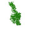
Mass: 24025.070 Da / Num. of mol.: 1 / Fragment: BAR DOMAIN, RESIDUES 1-143 Source method: isolated from a genetically manipulated source Source: (gene. exp.) RATTUS NORVEGICUS (Norway rat) / Production host: ESCHERICHIA COLI (E. coli) / Strain (production host): BL21 References: UniProt: O35179, Transferases; Acyltransferases; Transferring groups other than aminoacyl groups
Has protein modification
Y
Sequence details
UNIPROT DOES NOT CURRENTLY HAVE A FULL-LENGTH ENTRY FOR RAT ENDOPHILIN A1. THE FRAGMENTS FROM 25- ...UNIPROT DOES NOT CURRENTLY HAVE A FULL-LENGTH ENTRY FOR RAT ENDOPHILIN A1. THE FRAGMENTS FROM 25-104 HAS BEEN MAPPED TO ITSELF AT THE PRESENT TIME DUE TO A LACK OF AVAILABLE SEQUENCE DATABASE MAPPING
-
Experimental details
-
Experiment
Experiment
Method: X-RAY DIFFRACTION / Number of used crystals: 1
-
Sample preparation
Crystal
Density Matthews: 8.76 Å3/Da / Density % sol: 85.85 %
Crystal grow
Temperature: 277 K / pH: 8 Details: 25% BUTANE-1,4-DIOL, 100MM TRIS PH 8.0,4DEGC, CRYOPROTECTED IN 40% BUTANE-1,4-DIOL, 25% ETHYLENE GLYCOL
Resolution: 2.9→44 Å / Num. obs: 393336 / % possible obs: 99.9 % / Observed criterion σ(I): 6 / Redundancy: 22.1 % / Rmerge(I) obs: 0.16 / Net I/σ(I): 19.8
Reflection shell
Resolution: 2.9→3.06 Å / Redundancy: 22.3 % / Rmerge(I) obs: 0.95 / Mean I/σ(I) obs: 3.6 / % possible all: 100
-
Processing
Software
Name
Version
Classification
REFMAC
5.2.0005
refinement
MOSFLM
datareduction
SCALA
datascaling
SHARP
phasing
Refinement
Method to determine structure: MAD / Resolution: 2.9→89.44 Å / Cor.coef. Fo:Fc: 0.881 / Cor.coef. Fo:Fc free: 0.861 / SU B: 12.411 / SU ML: 0.228 / Cross valid method: THROUGHOUT / ESU R: 0.33 / ESU R Free: 0.273 Stereochemistry target values: MAXIMUM LIKELIHOODWITH PHASES Details: HYDROGENS HAVE BEEN ADDED IN THE RIDING POSITIONS.
Rfactor
Num. reflection
% reflection
Selection details
Rfree
0.295
904
5.1 %
RANDOM
Rwork
0.279
-
-
-
obs
0.28
16875
99.9 %
-
Solvent computation
Ion probe radii: 0.8 Å / Shrinkage radii: 0.8 Å / VDW probe radii: 1.2 Å / Solvent model: MASK
 Movie
Movie Controller


 Open data
 Basic information
 Components
 Keywords
 Function and homology information
 X-RAY DIFFRACTION /
  Authors
 Citation
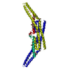
 Structure visualization
 Downloads & links
 Other downloads


 PDBj
PDBj


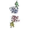
 Assembly
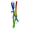

 Sample preparation
 / Beamline: ID29 / Wavelength: 0.98
  Processing