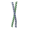
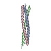
- PDB-1jcc: Crystal Structure of a Novel Alanine-Zipper Trimer at 1.7 A Resol... -
+
データを開く
IDまたはキーワード:
読み込み中...
-
基本情報
登録情報
データベース: PDB / ID: 1jcc
タイトル
Crystal Structure of a Novel Alanine-Zipper Trimer at 1.7 A Resolution, V13A,L16A,V20A,L23A,V27A,M30A,V34A mutations
要素
MAJOR OUTER MEMBRANE LIPOPROTEIN
キーワード
MEMBRANE PROTEIN / LIPOPROTEIN / PROTEIN FOLDING / COILED COIL / HELIX CAPPING / ALANINE-ZIPPER
機能・相同性
機能・相同性情報
periplasmic space organization / lipid modification / peptidoglycan binding / cell outer membrane / outer membrane-bounded periplasmic space / lipid binding / membrane / identical protein binding 類似検索 - 分子機能
Single alpha-helices involved in coiled-coils or other helix-helix interfaces - #190 / Lipoprotein leucine-zipper / Major outer membrane lipoprotein Lpp / Lipoprotein leucine-zipper / Single alpha-helices involved in coiled-coils or other helix-helix interfaces / Prokaryotic membrane lipoprotein lipid attachment site profile. / Up-down Bundle / Mainly Alpha 類似検索 - ドメイン・相同性
 ムービー
ムービー コントローラー


 データを開く
 基本情報
基本情報 要素
要素 キーワード
キーワード 機能・相同性情報
 X線回折 /
  データ登録者
 引用
引用 構造の表示
構造の表示 ダウンロードとリンク
 その他のダウンロード
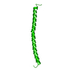


 PDBj
PDBj


 集合体
集合体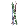

 分子量: 65.409 Da / 分子数: 3 / 由来タイプ: 合成 / 式: Zn
 分子量: 18.015 Da / 分子数: 234 / 由来タイプ: 天然 / 式: H2O
 試料調製
試料調製 / ビームライン: X26C / 波長: 1.1 / 波長: 1.1 Å
 解析
解析