 Movie
Movie Controller

[English] 日本語
 Yorodumi
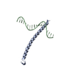
- PDB-2dgc: GCN4 BASIC DOMAIN, LEUCINE ZIPPER COMPLEXED WITH ATF/CREB SITE DNA -
+ Open data

- Basic information
| Entry | Database: PDB / ID: 2dgc | ||||||
|---|---|---|---|---|---|---|---|
| Title | GCN4 BASIC DOMAIN, LEUCINE ZIPPER COMPLEXED WITH ATF/CREB SITE DNA | ||||||
 Components Components |
| ||||||
 Keywords Keywords | TRANSCRIPTION/DNA / BASIC DOMAIN / LEUCINE ZIPPER / DNA BINDING / EUKARYOTIC REGULATORY PROTEIN / TRANSCRIPTION-DNA COMPLEX | ||||||
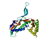
| Function / homology |  Function and homology information Function and homology informationFCERI mediated MAPK activation / protein localization to nuclear periphery / Activation of the AP-1 family of transcription factors / negative regulation of ribosomal protein gene transcription by RNA polymerase II / positive regulation of cellular response to amino acid starvation / response to amino acid starvation / mediator complex binding / Oxidative Stress Induced Senescence / amino acid biosynthetic process / TFIID-class transcription factor complex binding ...FCERI mediated MAPK activation / protein localization to nuclear periphery / Activation of the AP-1 family of transcription factors / negative regulation of ribosomal protein gene transcription by RNA polymerase II / positive regulation of cellular response to amino acid starvation / response to amino acid starvation / mediator complex binding / Oxidative Stress Induced Senescence / amino acid biosynthetic process / TFIID-class transcription factor complex binding / positive regulation of RNA polymerase II transcription preinitiation complex assembly / positive regulation of transcription initiation by RNA polymerase II / cellular response to nutrient levels / cellular response to amino acid starvation / RNA polymerase II transcription regulator complex / DNA-binding transcription activator activity, RNA polymerase II-specific / transcription regulator complex / sequence-specific DNA binding / RNA polymerase II-specific DNA-binding transcription factor binding / DNA-binding transcription factor activity, RNA polymerase II-specific / intracellular signal transduction / RNA polymerase II cis-regulatory region sequence-specific DNA binding / DNA-binding transcription factor activity / chromatin binding / negative regulation of transcription by RNA polymerase II / positive regulation of transcription by RNA polymerase II / identical protein binding / nucleus Similarity search - Function | ||||||
| Biological species |  | ||||||
| Method |  X-RAY DIFFRACTION / SYNCHROTRON / Resolution: 2.2 Å X-RAY DIFFRACTION / SYNCHROTRON / Resolution: 2.2 Å | ||||||
 Authors Authors | Keller, W. / Koenig, P. / Richmond, T.J. | ||||||
 Citation Citation | Journal: J.Mol.Biol. / Year: 1995 Title: Crystal structure of a bZIP/DNA complex at 2.2 A: determinants of DNA specific recognition. Authors: Keller, W. / Konig, P. / Richmond, T.J. #1: Journal: J.Mol.Biol. / Year: 1993Title: The X-Ray Structure of the GCN4-bZIP Bound to ATF/CREB Site DNA Shows the Complex Depends on DNA Flexibility Authors: Koenig, P. / Richmond, T.J. #2: Journal: Cell(Cambridge,Mass.) / Year: 1992Title: The GCN4 Basic Region Leucine Zipper Binds DNA as a Dimer of Uninterrupted Alpha Helices: Crystal Structure of the Protein-DNA Complex Authors: Ellenberger, T.E. / Brandl, C.J. / Struhl, K. / Harrison, S.C. | ||||||
| History |
|
- Structure visualization
| Structure viewer | Molecule: MolmilJmol/JSmol |
|---|
- Downloads & links
-Download
| PDBx/mmCIF format | 2dgc.cif.gz | 37.8 KB | Display | PDBx/mmCIF format |
|---|---|---|---|---|
| PDB format | pdb2dgc.ent.gz | 23 KB | Display | PDB format |
| PDBx/mmJSON format | 2dgc.json.gz | Tree view | PDBx/mmJSON format | |
| Others |  Other downloads Other downloads |
-Validation report
| Arichive directory | https://data.pdbj.org/pub/pdb/validation_reports/dg/2dgcftp://data.pdbj.org/pub/pdb/validation_reports/dg/2dgc | HTTPS FTP |
|---|
-Related structure data
| Related structure data | |
|---|---|
| Similar structure data |


-Links
 PDBj
PDBj


- Assembly
| Deposited unit | 
| ||||||||
|---|---|---|---|---|---|---|---|---|---|
| 1 | 
| ||||||||
| Unit cell |
| ||||||||
| Details | THE ASYMMETRIC UNIT CONTAINS ONE HALF OF PROTEIN/DNA COMPLEX PER ASYMMETRIC UNIT. MOLECULAR DYAD AXIS OF PROTEIN DIMER AND PALINDROMIC HALF SITES OF THE DNA COINCIDES WITH CRYSTALLOGRAPHIC TWO-FOLD AXIS. THE FULL PROTEIN/DNA COMPLEX CAN BE OBTAINED BY APPLYING THE FOLLOWING TRANSFORMATION MATRIX AND TRANSLATION VECTOR TO THE COORDINATES X Y Z: SYMMETRY THE CRYSTALLOGRAPHIC SYMMETRY TRANSFORMATIONS PRESENTED BELOW GENERATE THE SUBUNITS OF THE POLYMERIC MOLECULE. APPLIED TO RESIDUES: A 229 .. 277 APPLIED TO RESIDUES: B -10 .. 9 SYMMETRY1 1 0.000000 -1.000000 0.000000 117.32000 SYMMETRY2 1 -1.000000 0.000000 0.000000 117.32000 SYMMETRY3 1 0.000000 0.000000 -1.000000 43.44000 |
-Components
| #1: DNA chain | Mass: 5820.771 Da / Num. of mol.: 1 / Source method: obtained synthetically |
|---|---|
| #2: Protein | Mass: 7375.564 Da / Num. of mol.: 1 / Source method: isolated from a natural source / Source: (natural) |
| #3: Water | ChemComp-HOH /  Mass: 18.015 Da / Num. of mol.: 46 / Source method: isolated from a natural source / Formula: H2O Mass: 18.015 Da / Num. of mol.: 46 / Source method: isolated from a natural source / Formula: H2O |
| Sequence details | AMINO ACID NUMBERING (RESIDUE NUMBER) CORRESPONDS TO THE 281 AMINO ACIDS OF INTACT GCN4. RESIDUE ...AMINO ACID NUMBERING (RESIDUE NUMBER) CORRESPOND |
-Experimental details
-Experiment
| Experiment | Method: X-RAY DIFFRACTION / Number of used crystals: 1 |
|---|
- Sample preparation
| Crystal | Density Matthews: 2.78 Å3/Da / Density % sol: 55.82 % | ||||||||||||||||||||||||||||||||||||||||||||||||||||||||
|---|---|---|---|---|---|---|---|---|---|---|---|---|---|---|---|---|---|---|---|---|---|---|---|---|---|---|---|---|---|---|---|---|---|---|---|---|---|---|---|---|---|---|---|---|---|---|---|---|---|---|---|---|---|---|---|---|---|
| Crystal grow | Method: vapor diffusion / pH: 4.6 / Details: pH 4.60, VAPOR DIFFUSION | ||||||||||||||||||||||||||||||||||||||||||||||||||||||||
| Components of the solutions |
| ||||||||||||||||||||||||||||||||||||||||||||||||||||||||
| Crystal grow | *PLUS Method: unknown / pH: 4.6 | ||||||||||||||||||||||||||||||||||||||||||||||||||||||||
| Components of the solutions | *PLUS
|
-Data collection
| Diffraction | Mean temperature: 277 K |
|---|---|
| Diffraction source | Source: SYNCHROTRON / Site: EMBL/DESY, HAMBURG  / Beamline: X11 / Wavelength: 0.92 Å / Beamline: X11 / Wavelength: 0.92 Å |
| Detector | Type: MARRESEARCH / Detector: IMAGE PLATE |
| Radiation | Protocol: SINGLE WAVELENGTH / Monochromatic (M) / Laue (L): M / Scattering type: x-ray |
| Radiation wavelength | Wavelength: 0.92 Å / Relative weight: 1 |
| Reflection | Resolution: 2.2→15 Å / Num. obs: 6597 / % possible obs: 80.8 % / Observed criterion σ(F): 3 / Redundancy: 5 % / Rmerge(I) obs: 0.09 |
| Reflection | *PLUS Highest resolution: 2.2 Å / Lowest resolution: 15 Å / % possible obs: 80.8 % / Observed criterion σ(F): 3 |
- Processing
| Software |
| ||||||||||||||||||||||||||||||||||||||||||||||||||||||||||||
|---|---|---|---|---|---|---|---|---|---|---|---|---|---|---|---|---|---|---|---|---|---|---|---|---|---|---|---|---|---|---|---|---|---|---|---|---|---|---|---|---|---|---|---|---|---|---|---|---|---|---|---|---|---|---|---|---|---|---|---|---|---|
| Refinement | Resolution: 2.2→6 Å / σ(F): 3
| ||||||||||||||||||||||||||||||||||||||||||||||||||||||||||||
| Displacement parameters | Biso mean: 41.3 Å2 | ||||||||||||||||||||||||||||||||||||||||||||||||||||||||||||
| Refine analyze | Luzzati coordinate error obs: 0.32 Å | ||||||||||||||||||||||||||||||||||||||||||||||||||||||||||||
| Refinement step | Cycle: LAST / Resolution: 2.2→6 Å
| ||||||||||||||||||||||||||||||||||||||||||||||||||||||||||||
| Refine LS restraints |
| ||||||||||||||||||||||||||||||||||||||||||||||||||||||||||||
| Xplor file | Serial no: 1 / Param file: PARAM11.DNA | ||||||||||||||||||||||||||||||||||||||||||||||||||||||||||||
| Software | *PLUS Name: X-PLOR / Classification: refinement | ||||||||||||||||||||||||||||||||||||||||||||||||||||||||||||
| Refinement | *PLUS Highest resolution: 2.2 Å / Lowest resolution: 6 Å / σ(F): 3 | ||||||||||||||||||||||||||||||||||||||||||||||||||||||||||||
| Solvent computation | *PLUS | ||||||||||||||||||||||||||||||||||||||||||||||||||||||||||||
| Displacement parameters | *PLUS Biso mean: 41.3 Å2 |
