 Movie
Movie Controller


+ Open data

- Basic information
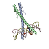
| Entry | Database: PDB / ID: 2c9l | ||||||
|---|---|---|---|---|---|---|---|
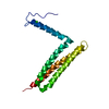
| Title | Structure of the Epstein-Barr virus ZEBRA protein | ||||||
 Components Components |
| ||||||
 Keywords Keywords | VIRAL PROTEIN / EPSTEIN-BARR VIRUS / EBV / ZEBRA / BZLF1 / ZTA / Z / LYTIC CYCLE ACTIVATION / BZIP PROTEIN / VIRAL PROTEIN DNA-BINDING / NUCLEAR PROTEIN / TRANSCRIPTION REGULATION | ||||||
| Function / homology |  Function and homology information Function and homology informationsymbiont-mediated suppression of host tumor necrosis factor-mediated signaling pathway / symbiont-mediated perturbation of host cell cycle G0/G1 transition checkpoint / release from viral latency / symbiont-mediated suppression of host cytoplasmic pattern recognition receptor signaling pathway via inhibition of IRF7 activity / symbiont-mediated perturbation of host cell cycle G1/S transition checkpoint / sequence-specific DNA binding / protein dimerization activity / DNA-binding transcription factor activity / regulation of DNA-templated transcription / positive regulation of DNA-templated transcription ...symbiont-mediated suppression of host tumor necrosis factor-mediated signaling pathway / symbiont-mediated perturbation of host cell cycle G0/G1 transition checkpoint / release from viral latency / symbiont-mediated suppression of host cytoplasmic pattern recognition receptor signaling pathway via inhibition of IRF7 activity / symbiont-mediated perturbation of host cell cycle G1/S transition checkpoint / sequence-specific DNA binding / protein dimerization activity / DNA-binding transcription factor activity / regulation of DNA-templated transcription / positive regulation of DNA-templated transcription / chromatin / host cell nucleus / DNA binding Similarity search - Function | ||||||
| Biological species |  HUMAN HERPESVIRUS 4 (Epstein-Barr virus) HUMAN HERPESVIRUS 4 (Epstein-Barr virus) | ||||||
| Method |  X-RAY DIFFRACTION / SYNCHROTRON / MOLECULAR REPLACEMENT / Resolution: 2.25 Å X-RAY DIFFRACTION / SYNCHROTRON / MOLECULAR REPLACEMENT / Resolution: 2.25 Å | ||||||
 Authors Authors | Petosa, C. / Morand, P. / Baudin, F. / Moulin, M. / Artero, J.B. / Muller, C.W. | ||||||
 Citation Citation | Journal: Mol.Cell / Year: 2006 Title: Structural Basis of Lytic Cycle Activation by the Epstein-Barr Virus Zebra Protein Authors: Petosa, C. / Morand, P. / Baudin, F. / Moulin, M. / Artero, J.B. / Muller, C.W. | ||||||
| History |
|
- Structure visualization
| Structure viewer | Molecule: MolmilJmol/JSmol |
|---|
- Downloads & links
-Download
| PDBx/mmCIF format | 2c9l.cif.gz | 62.8 KB | Display | PDBx/mmCIF format |
|---|---|---|---|---|
| PDB format | pdb2c9l.ent.gz | 43.3 KB | Display | PDB format |
| PDBx/mmJSON format | 2c9l.json.gz | Tree view | PDBx/mmJSON format | |
| Others |  Other downloads Other downloads |
-Validation report
| Arichive directory | https://data.pdbj.org/pub/pdb/validation_reports/c9/2c9lftp://data.pdbj.org/pub/pdb/validation_reports/c9/2c9l | HTTPS FTP |
|---|
-Related structure data
| Related structure data | 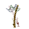 2c9nC 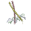 1ysaS C: citing same article ( S: Starting model for refinement |
|---|---|
| Similar structure data |


-Links
 PDBj
PDBj


- Assembly
| Deposited unit | 
| ||||||||
|---|---|---|---|---|---|---|---|---|---|
| 1 |
| ||||||||
| Unit cell |
|
-Components
| #1: DNA chain | Mass: 5837.812 Da / Num. of mol.: 1 / Source method: obtained synthetically / Source: (synth.) HUMAN HERPESVIRUS 4 (Epstein-Barr virus) | ||||
|---|---|---|---|---|---|
| #2: DNA chain | Mass: 5506.577 Da / Num. of mol.: 1 / Source method: obtained synthetically / Source: (synth.) HUMAN HERPESVIRUS 4 (Epstein-Barr virus) | ||||
| #3: Protein | Mass: 7413.740 Da / Num. of mol.: 2 Fragment: DNA-BINDING AND DIMERIZATION DOMAIN, RESIDUES 175-236 Mutation: YES Source method: isolated from a genetically manipulated source Source: (gene. exp.) HUMAN HERPESVIRUS 4 (Epstein-Barr virus)Production host:  #4: Water | ChemComp-HOH / |  Mass: 18.015 Da / Num. of mol.: 81 / Source method: isolated from a natural source / Formula: H2O Mass: 18.015 Da / Num. of mol.: 81 / Source method: isolated from a natural source / Formula: H2OCompound details | ENGINEERED RESIDUE IN CHAIN Y, SER 186 TO ALA ENGINEERED RESIDUE IN CHAIN Z, CYS 189 TO SER ...ENGINEERED | |
-Experimental details
-Experiment
| Experiment | Method: X-RAY DIFFRACTION / Number of used crystals: 1 |
|---|
- Sample preparation
| Crystal | Density Matthews: 2.26 Å3/Da / Density % sol: 45.16 % |
|---|---|
| Crystal grow | pH: 7 / Details: pH 7.00 |
-Data collection
| Diffraction | Mean temperature: 100 K |
|---|---|
| Diffraction source | Source: SYNCHROTRON / Site: ESRF  / Beamline: ID13 / Wavelength: 0.9755 / Beamline: ID13 / Wavelength: 0.9755 |
| Detector | Type: MARRESEARCH / Detector: CCD / Date: Sep 11, 2004 |
| Radiation | Protocol: SINGLE WAVELENGTH / Monochromatic (M) / Laue (L): M / Scattering type: x-ray |
| Radiation wavelength | Wavelength: 0.9755 Å / Relative weight: 1 |
| Reflection | Resolution: 2.25→30 Å / Num. obs: 11609 / % possible obs: 99.8 % / Observed criterion σ(I): 0 / Redundancy: 5.3 % / Rmerge(I) obs: 0.09 / Net I/σ(I): 11.1 |
| Reflection shell | Resolution: 2.25→2.3 Å / Redundancy: 5.4 % / Rmerge(I) obs: 0.45 / Mean I/σ(I) obs: 4.4 / % possible all: 99.9 |
- Processing
| Software |
| ||||||||||||||||||||||||||||||||||||||||||||||||||||||||||||
|---|---|---|---|---|---|---|---|---|---|---|---|---|---|---|---|---|---|---|---|---|---|---|---|---|---|---|---|---|---|---|---|---|---|---|---|---|---|---|---|---|---|---|---|---|---|---|---|---|---|---|---|---|---|---|---|---|---|---|---|---|---|
| Refinement | Method to determine structure: MOLECULAR REPLACEMENT Starting model: PDB ENTRY 1YSA Resolution: 2.25→30 Å / Data cutoff high absF: 10000 / Cross valid method: THROUGHOUT / σ(F): 0 Details: THERE IS NO SIDE CHAIN DENSITY VISIBLE FOR RESIDUES LEU175 AND GLU176 IN CHAIN Z. ONLY THE MAIN CHAIN ATOMS OF THESE RESIDUES ARE INCLUDED IN THE MODEL.
| ||||||||||||||||||||||||||||||||||||||||||||||||||||||||||||
| Solvent computation | Bsol: 56.8112 Å2 / ksol: 0.335561 e/Å3 | ||||||||||||||||||||||||||||||||||||||||||||||||||||||||||||
| Displacement parameters |
| ||||||||||||||||||||||||||||||||||||||||||||||||||||||||||||
| Refinement step | Cycle: LAST / Resolution: 2.25→30 Å
| ||||||||||||||||||||||||||||||||||||||||||||||||||||||||||||
| Refine LS restraints |
| ||||||||||||||||||||||||||||||||||||||||||||||||||||||||||||
| Xplor file |
|
