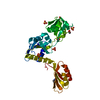
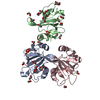
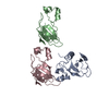
[English] 日本語
Yorodumi
- PDB-1k04: Crystal Structure of the Focal Adhesion Targeting Domain of Focal... -

+
Open data


ID or keywords:

Loading...

-
Basic information

Entry
Database: PDB / ID: 1k04
TitleCrystal Structure of the Focal Adhesion Targeting Domain of Focal Adhesion Kinase
ComponentsFOCAL ADHESION KINASE 1PTK2
KeywordsTRANSFERASE / up-down-up-down four helical bundle forming a helix-exchange dimer
Function / homology
Function and homology information


netrin-activated signaling pathway / regulation of substrate adhesion-dependent cell spreading / regulation of endothelial cell migration / regulation of epithelial cell migration / detection of muscle stretch / positive regulation of ubiquitin-dependent protein catabolic process / JUN kinase binding / signal complex assembly / positive regulation of macrophage proliferation / positive regulation of fibroblast migration ...netrin-activated signaling pathway / regulation of substrate adhesion-dependent cell spreading / regulation of endothelial cell migration / regulation of epithelial cell migration / detection of muscle stretch / positive regulation of ubiquitin-dependent protein catabolic process / JUN kinase binding / signal complex assembly / positive regulation of macrophage proliferation / positive regulation of fibroblast migration / DCC mediated attractive signaling / Signal regulatory protein family interactions / growth hormone receptor signaling pathway / regulation of osteoblast differentiation / regulation of cytoskeleton organization / MET activates PTK2 signaling / regulation of focal adhesion assembly / establishment of cell polarity / positive regulation of wound healing / positive regulation of macrophage chemotaxis / regulation of GTPase activity / Fc-gamma receptor signaling pathway involved in phagocytosis / p130Cas linkage to MAPK signaling for integrins / positive regulation of epithelial cell migration / negative regulation of cell-cell adhesion / Apoptotic cleavage of cellular proteins / regulation of cell adhesion mediated by integrin / GRB2:SOS provides linkage to MAPK signaling for Integrins / negative regulation of anoikis / Estrogen-dependent nuclear events downstream of ESR-membrane signaling / RHO GTPases Activate WASPs and WAVEs / positive regulation of epithelial to mesenchymal transition / ephrin receptor signaling pathway / positive regulation of protein kinase activity / vascular endothelial growth factor receptor signaling pathway / regulation of cell adhesion / heart morphogenesis / stress fiber / EPHB-mediated forward signaling / NCAM signaling for neurite out-growth / SH2 domain binding / Integrin signaling / transforming growth factor beta receptor signaling pathway / ciliary basal body / placenta development / protein tyrosine phosphatase activity / molecular function activator activity / cell motility / integrin-mediated signaling pathway / axon guidance / FCGR3A-mediated phagocytosis / regulation of protein phosphorylation / non-specific protein-tyrosine kinase / non-membrane spanning protein tyrosine kinase activity / epidermal growth factor receptor signaling pathway / Regulation of actin dynamics for phagocytic cup formation / VEGFA-VEGFR2 Pathway / peptidyl-tyrosine phosphorylation / cell migration / integrin binding / actin binding / regulation of cell population proliferation / cell cortex / regulation of cell shape / RAF/MAP kinase cascade / protein phosphatase binding / angiogenesis / protein tyrosine kinase activity / dendritic spine / Extra-nuclear estrogen signaling / protein autophosphorylation / positive regulation of phosphatidylinositol 3-kinase/protein kinase B signal transduction / cytoskeleton / positive regulation of cell migration / positive regulation of protein phosphorylation / focal adhesion / intracellular membrane-bounded organelle / centrosome / positive regulation of cell population proliferation / negative regulation of apoptotic process / protein kinase binding / perinuclear region of cytoplasm / ATP binding / nucleus / plasma membrane / cytosol / cytoplasm
Similarity search - Function
Single helix bin / Nucleotidyltransferases domain 2 / Focal adhesion kinase, targeting (FAT) domain / Focal adhesion kinase, targeting (FAT) domain superfamily / Focal adhesion kinase, N-terminal / FAK1/PYK2, FERM domain C-lobe / Focal adhesion targeting region / FERM N-terminal domain / : / FAK1/PYK2, FERM domain C-lobe ...Single helix bin / Nucleotidyltransferases domain 2 / Focal adhesion kinase, targeting (FAT) domain / Focal adhesion kinase, targeting (FAT) domain superfamily / Focal adhesion kinase, N-terminal / FAK1/PYK2, FERM domain C-lobe / Focal adhesion targeting region / FERM N-terminal domain / : / FAK1/PYK2, FERM domain C-lobe / FERM domain signature 2. / FERM central domain / FERM/acyl-CoA-binding protein superfamily / FERM central domain / FERM superfamily, second domain / FERM domain / FERM domain profile. / Band 4.1 domain / Band 4.1 homologues / Four Helix Bundle (Hemerythrin (Met), subunit A) / Single alpha-helices involved in coiled-coils or other helix-helix interfaces / Tyrosine-protein kinase, catalytic domain / Tyrosine kinase, catalytic domain / Tyrosine protein kinases specific active-site signature. / PH-like domain superfamily / Tyrosine-protein kinase, active site / Protein tyrosine and serine/threonine kinase / Serine-threonine/tyrosine-protein kinase, catalytic domain / Ubiquitin-like domain superfamily / Protein kinase, ATP binding site / Protein kinases ATP-binding region signature. / Protein kinase domain profile. / Protein kinase domain / Protein kinase-like domain superfamily / Up-down Bundle / Mainly Alpha
Similarity search - Domain/homology
Focal adhesion kinase 1
Similarity search - Component
Biological speciesHomo sapiens (human)
MethodX-RAY DIFFRACTION / SYNCHROTRON / MOLECULAR REPLACEMENT / Resolution: 1.95 Å
AuthorsArold, S.T. / Hoellerer, M.K. / Noble, M.E.M.
CitationJournal: Structure / Year: 2002
Title: The structural basis of localization and signaling by the focal adhesion targeting domain.
Authors: Arold, S.T. / Hoellerer, M.K. / Noble, M.E.
History
DepositionSep 18, 2001Deposition site: RCSB / Processing site: RCSB
Revision 1.0Jan 30, 2002Provider: repository / Type: Initial release
Revision 1.1Apr 27, 2008Group: Version format compliance
Revision 1.2Jul 13, 2011Group: Derived calculations / Version format compliance
Revision 1.3Feb 7, 2024Group: Data collection / Database references / Derived calculations
Category: chem_comp_atom / chem_comp_bond ...chem_comp_atom / chem_comp_bond / database_2 / pdbx_struct_special_symmetry / struct_site
Item: _database_2.pdbx_DOI / _database_2.pdbx_database_accession ..._database_2.pdbx_DOI / _database_2.pdbx_database_accession / _struct_site.pdbx_auth_asym_id / _struct_site.pdbx_auth_comp_id / _struct_site.pdbx_auth_seq_id

-
Structure visualization

Structure viewerMolecule:
MolmilJmol/JSmol

Downloads & links

-
Assembly

Deposited unit
A: FOCAL ADHESION KINASE 1
hetero molecules


Theoretical massNumber of molelcules
Total (without water)17,9852
Polymers17,9501
Non-polymers351
Water1,74797
1
A: FOCAL ADHESION KINASE 1
hetero molecules

A: FOCAL ADHESION KINASE 1
hetero molecules


Theoretical massNumber of molelcules
Total (without water)35,9704
Polymers35,8992
Non-polymers712
Water362
TypeNameSymmetry operationNumber
identity operation1_555x,y,z1
crystal symmetry operation3_655-x+1,y,-z+1/21
Buried area4710 Å2
ΔGint-52 kcal/mol
Surface area16940 Å2
MethodPISA, PQS
Unit cell
Length a, b, c (Å)38.426, 95.555, 84.821
Angle α, β, γ (deg.)90.00, 90.00, 90.00
Int Tables number20
Space group name H-MC2221
Components on special symmetry positions
IDModelComponents
11A-200-

CL

21A-1-

HOH

31A-52-

HOH

41A-107-

HOH

DetailsThe second protomer of the dimeric assembly is generated by the two fold axis: -X,-Y,1/2+Z

-
Components

#1: Protein FOCAL ADHESION KINASE 1 / PTK2 / FADK 1


Mass: 17949.697 Da / Num. of mol.: 1
Source method: isolated from a genetically manipulated source
Source: (gene. exp.) Homo sapiens (human) / Gene: FAK / Plasmid: pGEX-6P2 / Production host: Escherichia coli (E. coli) / Strain (production host): B834(DE3)pLysS / References: UniProt: Q05397, EC: 2.7.1.112
#2: Chemical ChemComp-CL / CHLORIDE ION / Chloride


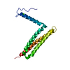
Mass: 35.453 Da / Num. of mol.: 1 / Source method: obtained synthetically / Formula: Cl
#3: Water ChemComp-HOH / water / Water


Mass: 18.015 Da / Num. of mol.: 97 / Source method: isolated from a natural source / Formula: H2O

-
Experimental details

-
Experiment

ExperimentMethod: X-RAY DIFFRACTION / Number of used crystals: 1

-
Sample preparation

CrystalDensity Matthews: 2.17 Å3/Da / Density % sol: 42.9 %
Crystal growTemperature: 296 K / Method: vapor diffusion, hanging drop / pH: 6.5
Details: MES, NaCl, Na/K phosphate, pH 6.5, VAPOR DIFFUSION, HANGING DROP, temperature 296K
Crystal grow
*PLUS
pH: 8
Components of the solutions
*PLUS
IDConc.Common nameCrystal-IDSol-IDDetailsChemical formula
110 mMTris-HCl1droppH8.0
2150 mM1dropNaCl
32 mMEDTA1drop
42 mMdithiothreitol1drop
520 mg/mlprotein1drop
60.1 MMES1reservoirpH6.5
72.0 M1reservoirNaCl
80.2 Msodium potassium phosphate1reservoir

-
Data collection

DiffractionMean temperature: 100 K
Diffraction sourceSource: SYNCHROTRON / Site: ESRF / Beamline: ID14-3 / Wavelength: 0.931 Å
DetectorType: ADSC QUANTUM 4 / Detector: CCD / Date: Nov 3, 2000
RadiationProtocol: SINGLE WAVELENGTH / Monochromatic (M) / Laue (L): M / Scattering type: x-ray
Radiation wavelengthWavelength: 0.931 Å / Relative weight: 1
ReflectionResolution: 1.95→47.7 Å / Num. obs: 11110 / % possible obs: 94.8 % / Observed criterion σ(F): 0.9 / Observed criterion σ(I): 0.9 / Redundancy: 3.3 % / Biso Wilson estimate: 45.7 Å2 / Rsym value: 0.056 / Net I/σ(I): 2.4
Reflection shellResolution: 1.95→2.02 Å / Redundancy: 2.5 % / Mean I/σ(I) obs: 1.4 / Num. unique all: 2462 / Rsym value: 0.509 / % possible all: 88.9
Reflection
*PLUS
Num. measured all: 36960 / Rmerge(I) obs: 0.056
Reflection shell
*PLUS
% possible obs: 88.9 % / Rmerge(I) obs: 0.509

-
Processing

Software
NameVersionClassification
AMoREphasing
REFMACrefinement
MOSFLMdata reduction
CCP4(SCALA)data scaling
RefinementMethod to determine structure: MOLECULAR REPLACEMENT / Resolution: 1.95→47.7 Å / σ(F): 0 / σ(I): 0 / Stereochemistry target values: CNS
RfactorNum. reflection% reflectionSelection details
Rfree0.27 719 7.2 %random
Rwork0.243 ---
all-11110 --
obs-11110 94.8 %-
Displacement parameters
Baniso -1Baniso -2Baniso -3
1--19.527 Å20 Å20 Å2
2--12.764 Å20 Å2
3---6.763 Å2
Refinement stepCycle: LAST / Resolution: 1.95→47.7 Å
ProteinNucleic acidLigandSolventTotal
Num. atoms1098 0 1 97 1196
Refine LS restraints
Refine-IDTypeDev ideal
X-RAY DIFFRACTIONp_bond_d0.001
X-RAY DIFFRACTIONp_angle_d1.4
LS refinement shellResolution: 1.95→2.02 Å
RfactorNum. reflection% reflection
Rfree0.43 51 -
Rwork0.44 --
obs-999 8.5 %
Xplor file
Refine-IDSerial noParam file
X-RAY DIFFRACTION1protein_rep.param
X-RAY DIFFRACTION2water_rep.param
X-RAY DIFFRACTION3ion.param
Refinement
*PLUS
% reflection Rfree: 7 % / Rfactor obs: 0.243 / Rfactor Rfree: 0.27 / Rfactor Rwork: 0.243
Solvent computation
*PLUS
Displacement parameters
*PLUS
Refine LS restraints
*PLUS
Refine-IDTypeDev ideal
X-RAY DIFFRACTIONp_bond_d0.01
X-RAY DIFFRACTIONp_angle_d
X-RAY DIFFRACTIONp_angle_deg1.4
LS refinement shell
*PLUS
Rfactor Rfree: 0.43 / Rfactor Rwork: 0.44

+
About Yorodumi

-
News

-
Feb 9, 2022. New format data for meta-information of EMDB entries

New format data for meta-information of EMDB entries

  • Version 3 of the EMDB header file is now the official format.
  • The previous official version 1.9 will be removed from the archive.

Related info.:EMDB header

External links:wwPDB to switch to version 3 of the EMDB data model

-
Aug 12, 2020. Covid-19 info

Covid-19 info

URL: https://pdbj.org/emnavi/covid19.php

New page: Covid-19 featured information page in EM Navigator.

Related info.:Covid-19 info / Mar 5, 2020. Novel coronavirus structure data

+
Mar 5, 2020. Novel coronavirus structure data

Novel coronavirus structure data

Related info.:Yorodumi Speices / Aug 12, 2020. Covid-19 info

External links:COVID-19 featured content - PDBj / Molecule of the Month (242):Coronavirus Proteases

+
Jan 31, 2019. EMDB accession codes are about to change! (news from PDBe EMDB page)

EMDB accession codes are about to change! (news from PDBe EMDB page)

  • The allocation of 4 digits for EMDB accession codes will soon come to an end. Whilst these codes will remain in use, new EMDB accession codes will include an additional digit and will expand incrementally as the available range of codes is exhausted. The current 4-digit format prefixed with “EMD-” (i.e. EMD-XXXX) will advance to a 5-digit format (i.e. EMD-XXXXX), and so on. It is currently estimated that the 4-digit codes will be depleted around Spring 2019, at which point the 5-digit format will come into force.
  • The EM Navigator/Yorodumi systems omit the EMD- prefix.

Related info.:Q: What is EMD? / ID/Accession-code notation in Yorodumi/EM Navigator

External links:EMDB Accession Codes are Changing Soon! / Contact to PDBj

+
Jul 12, 2017. Major update of PDB

Major update of PDB

  • wwPDB released updated PDB data conforming to the new PDBx/mmCIF dictionary.
  • This is a major update changing the version number from 4 to 5, and with Remediation, in which all the entries are updated.
  • In this update, many items about electron microscopy experimental information are reorganized (e.g. em_software).
  • Now, EM Navigator and Yorodumi are based on the updated data.

External links:wwPDB Remediation / Enriched Model Files Conforming to OneDep Data Standards Now Available in the PDB FTP Archive

-
Yorodumi

Thousand views of thousand structures

  • Yorodumi is a browser for structure data from EMDB, PDB, SASBDB, etc.
  • This page is also the successor to EM Navigator detail page, and also detail information page/front-end page for Omokage search.
  • The word "yorodu" (or yorozu) is an old Japanese word meaning "ten thousand". "mi" (miru) is to see.

Related info.:EMDB / PDB / SASBDB / Comparison of 3 databanks / Yorodumi Search / Aug 31, 2016. New EM Navigator & Yorodumi / Yorodumi Papers / Jmol/JSmol / Function and homology information / Changes in new EM Navigator and Yorodumi

Read more