 Movie
Movie Controller

[English] 日本語
 Yorodumi
- PDB-5lht: ATP Phosphoribosyltransferase from Mycobacterium tuberculosis in ... -
+ Open data

- Basic information
| Entry | Database: PDB / ID: 5lht | ||||||
|---|---|---|---|---|---|---|---|
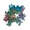
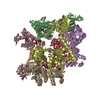
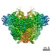
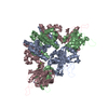
| Title | ATP Phosphoribosyltransferase from Mycobacterium tuberculosis in complex with the allosteric activator 3-(2-Thienyl)-L-alanine | ||||||
 Components Components | ATP phosphoribosyltransferase | ||||||
 Keywords Keywords | TRANSFERASE / ATP-PRTase / ACT / His G / Histidine biosynthesis | ||||||
| Function / homology |  Function and homology information Function and homology informationATP phosphoribosyltransferase / ATP phosphoribosyltransferase activity / L-histidine biosynthetic process / AMP binding / magnesium ion binding / ATP binding / plasma membrane / cytoplasm Similarity search - Function | ||||||
| Biological species |   Mycobacterium tuberculosis (bacteria) Mycobacterium tuberculosis (bacteria) | ||||||
| Method |  X-RAY DIFFRACTION / SYNCHROTRON / MOLECULAR REPLACEMENT / Resolution: 2.0601 Å X-RAY DIFFRACTION / SYNCHROTRON / MOLECULAR REPLACEMENT / Resolution: 2.0601 Å | ||||||
 Authors Authors | de Chiara, C. / Pisco, J.P. / de Carvalho, L.P. / Smerdon, S.J. / Walker, P.A. / Ogrodowicz, R. | ||||||
 Citation Citation | Journal: Nat Commun / Year: 2017 Title: Uncoupling conformational states from activity in an allosteric enzyme. Authors: Pisco, J.P. / Chiara, C. / Pacholarz, K.J. / Garza-Garcia, A. / Ogrodowicz, R.W. / Walker, P.A. / Barran, P.E. / Smerdon, S.J. / Carvalho, L.P.S. #1: Journal: Biochemistry / Year: 2012 Title: Mechanism of feedback allosteric inhibition of ATP phosphoribosyltransferase. Authors: Pedreno, S. / Pisco, J.P. / Larrouy-Maumus, G. / Kelly, G. / de Carvalho, L.P. | ||||||
| History |
|
- Structure visualization
| Structure viewer | Molecule: MolmilJmol/JSmol |
|---|
- Downloads & links
-Download
| PDBx/mmCIF format | 5lht.cif.gz | 71.8 KB | Display | PDBx/mmCIF format |
|---|---|---|---|---|
| PDB format | pdb5lht.ent.gz | 52.1 KB | Display | PDB format |
| PDBx/mmJSON format | 5lht.json.gz | Tree view | PDBx/mmJSON format | |
| Others |  Other downloads Other downloads |
-Validation report
| Arichive directory | https://data.pdbj.org/pub/pdb/validation_reports/lh/5lhtftp://data.pdbj.org/pub/pdb/validation_reports/lh/5lht | HTTPS FTP |
|---|
-Related structure data
| Related structure data | 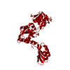 5lhuC  1nh8S S: Starting model for refinement C: citing same article ( |
|---|---|
| Similar structure data |


-Links
 PDBj
PDBj- Assembly
| Deposited unit | 
| ||||||||
|---|---|---|---|---|---|---|---|---|---|
| 1 | x 6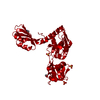
| ||||||||
| Unit cell |
|
-Components
| #1: Protein | Mass: 31686.023 Da / Num. of mol.: 1 Source method: isolated from a genetically manipulated source Source: (gene. exp.) Mycobacterium tuberculosis (strain ATCC 25618 / H37Rv) (bacteria)Gene: hisG, Rv2121c, MTCY261.17c / Production host: | ||||||
|---|---|---|---|---|---|---|---|
| #2: Chemical | ChemComp-TIH /   Type: L-peptide linking / Mass: 171.217 Da / Num. of mol.: 1 / Source method: obtained synthetically / Formula: C7H9NO2S Type: L-peptide linking / Mass: 171.217 Da / Num. of mol.: 1 / Source method: obtained synthetically / Formula: C7H9NO2S | ||||||
| #3: Chemical | ChemComp-SO4 /   Mass: 96.063 Da / Num. of mol.: 4 / Source method: obtained synthetically / Formula: SO4 Mass: 96.063 Da / Num. of mol.: 4 / Source method: obtained synthetically / Formula: SO4#4: Chemical | ChemComp-GOL / |   Mass: 92.094 Da / Num. of mol.: 1 / Source method: obtained synthetically / Formula: C3H8O3 Mass: 92.094 Da / Num. of mol.: 1 / Source method: obtained synthetically / Formula: C3H8O3#5: Water | ChemComp-HOH / |  Mass: 18.015 Da / Num. of mol.: 82 / Source method: isolated from a natural source / Formula: H2O Mass: 18.015 Da / Num. of mol.: 82 / Source method: isolated from a natural source / Formula: H2OHas protein modification | Y | |
-Experimental details
-Experiment
| Experiment | Method: X-RAY DIFFRACTION / Number of used crystals: 2 |
|---|
- Sample preparation
| Crystal |
| ||||||||||||
|---|---|---|---|---|---|---|---|---|---|---|---|---|---|
| Crystal grow |
|
-Data collection
| Diffraction | Mean temperature: 100 K |
|---|---|
| Diffraction source | Source: SYNCHROTRON / Site: Diamond  / Beamline: I04 / Wavelength: 0.9282 Å / Beamline: I04 / Wavelength: 0.9282 Å |
| Detector | Type: DECTRIS PILATUS 6M-F / Detector: PIXEL / Date: Dec 6, 2015 |
| Radiation | Protocol: SINGLE WAVELENGTH / Monochromatic (M) / Laue (L): M / Scattering type: x-ray |
| Radiation wavelength | Wavelength: 0.9282 Å / Relative weight: 1 |
| Reflection | Resolution: 2.0609→47.2522 Å / Num. obs: 20991 / % possible obs: 99.7 % / Redundancy: 4.6 % / Rmerge(I) obs: 0.041 / Net I/σ(I): 16.1 |
| Reflection shell | Rmerge(I) obs: 0.69 |
- Processing
| Software |
| ||||||||||||||||||||||||||||||||||||||||||||||||||||||||
|---|---|---|---|---|---|---|---|---|---|---|---|---|---|---|---|---|---|---|---|---|---|---|---|---|---|---|---|---|---|---|---|---|---|---|---|---|---|---|---|---|---|---|---|---|---|---|---|---|---|---|---|---|---|---|---|---|---|
| Refinement | Method to determine structure: MOLECULAR REPLACEMENT Starting model: 1NH8 Resolution: 2.0601→47.2522 Å / SU ML: 0.24 / Cross valid method: FREE R-VALUE / σ(F): 1.34 / Phase error: 25.37 / Stereochemistry target values: ML
| ||||||||||||||||||||||||||||||||||||||||||||||||||||||||
| Solvent computation | Shrinkage radii: 0.9 Å / VDW probe radii: 1.11 Å / Solvent model: FLAT BULK SOLVENT MODEL | ||||||||||||||||||||||||||||||||||||||||||||||||||||||||
| Refinement step | Cycle: LAST / Resolution: 2.0601→47.2522 Å
| ||||||||||||||||||||||||||||||||||||||||||||||||||||||||
| Refine LS restraints |
| ||||||||||||||||||||||||||||||||||||||||||||||||||||||||
| LS refinement shell |
|
