 Movie
Movie Controller

[English] 日本語
 Yorodumi
- PDB-1k05: Crystal structure of the Focal Adhesion Targeting Domain of Focal... -
+ Open data

- Basic information
| Entry | Database: PDB / ID: 1k05 | ||||||
|---|---|---|---|---|---|---|---|
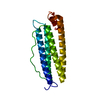
| Title | Crystal structure of the Focal Adhesion Targeting Domain of Focal Adhesion Kinase | ||||||
 Components Components | FOCAL ADHESION KINASE 1 | ||||||
 Keywords Keywords | TRANSFERASE / up-down-up-down four helical bundle | ||||||
| Function / homology |  Function and homology information Function and homology informationnetrin-activated signaling pathway / regulation of substrate adhesion-dependent cell spreading / regulation of endothelial cell migration / regulation of epithelial cell migration / detection of muscle stretch / positive regulation of cell-cell adhesion mediated by integrin / JUN kinase binding / signal complex assembly / negative regulation of cell adhesion mediated by integrin / positive regulation of macrophage proliferation ...netrin-activated signaling pathway / regulation of substrate adhesion-dependent cell spreading / regulation of endothelial cell migration / regulation of epithelial cell migration / detection of muscle stretch / positive regulation of cell-cell adhesion mediated by integrin / JUN kinase binding / signal complex assembly / negative regulation of cell adhesion mediated by integrin / positive regulation of macrophage proliferation / DCC mediated attractive signaling / Signal regulatory protein family interactions / positive regulation of fibroblast migration / MET activates PTK2 signaling / growth hormone receptor signaling pathway / positive regulation of ubiquitin-dependent protein catabolic process / regulation of focal adhesion assembly / negative regulation of cell-cell adhesion / Apoptotic cleavage of cellular proteins / p130Cas linkage to MAPK signaling for integrins / positive regulation of wound healing / regulation of osteoblast differentiation / Fc-gamma receptor signaling pathway involved in phagocytosis / regulation of cytoskeleton organization / positive regulation of macrophage chemotaxis / establishment of cell polarity / vascular endothelial cell response to oscillatory fluid shear stress / GRB2:SOS provides linkage to MAPK signaling for Integrins / negative regulation of anoikis / positive regulation of epithelial cell migration / vascular endothelial growth factor receptor signaling pathway / Estrogen-dependent nuclear events downstream of ESR-membrane signaling / ephrin receptor signaling pathway / RHO GTPases Activate WASPs and WAVEs / positive regulation of epithelial to mesenchymal transition / regulation of cell adhesion / heart morphogenesis / Integrin signaling / stress fiber / EPHB-mediated forward signaling / protein tyrosine phosphatase activity / transforming growth factor beta receptor signaling pathway / NCAM signaling for neurite out-growth / SH2 domain binding / placenta development / molecular function activator activity / Turbulent (oscillatory, disturbed) flow shear stress activates signaling by PIEZO1 and integrins in endothelial cells / axon guidance / ciliary tip / integrin-mediated signaling pathway / non-specific protein-tyrosine kinase / FCGR3A-mediated phagocytosis / cell motility / non-membrane spanning protein tyrosine kinase activity / Regulation of actin dynamics for phagocytic cup formation / VEGFA-VEGFR2 Pathway / integrin binding / epidermal growth factor receptor signaling pathway / regulation of cell shape / regulation of cell population proliferation / cell migration / RAF/MAP kinase cascade / actin binding / protein tyrosine kinase activity / angiogenesis / protein phosphatase binding / cell cortex / cytoskeleton / positive regulation of phosphatidylinositol 3-kinase/protein kinase B signal transduction / Extra-nuclear estrogen signaling / cilium / positive regulation of cell migration / ciliary basal body / focal adhesion / positive regulation of cell population proliferation / centrosome / protein kinase binding / negative regulation of apoptotic process / perinuclear region of cytoplasm / ATP binding / nucleus / plasma membrane / cytosol Similarity search - Function | ||||||
| Biological species |  Homo sapiens (human) Homo sapiens (human) | ||||||
| Method |  X-RAY DIFFRACTION / SYNCHROTRON / MIRAS / Resolution: 2.9 Å X-RAY DIFFRACTION / SYNCHROTRON / MIRAS / Resolution: 2.9 Å | ||||||
 Authors Authors | Arold, S.T. / Hoellerer, M.K. / Noble, M.E.M. | ||||||
 Citation Citation | Journal: Structure / Year: 2002 Title: The structural basis of localization and signaling by the focal adhesion targeting domain. Authors: Arold, S.T. / Hoellerer, M.K. / Noble, M.E. | ||||||
| History |
|
- Structure visualization
| Structure viewer | Molecule: MolmilJmol/JSmol |
|---|
- Downloads & links
-Download
| PDBx/mmCIF format | 1k05.cif.gz | 88.8 KB | Display | PDBx/mmCIF format |
|---|---|---|---|---|
| PDB format | pdb1k05.ent.gz | 70.1 KB | Display | PDB format |
| PDBx/mmJSON format | 1k05.json.gz | Tree view | PDBx/mmJSON format | |
| Others |  Other downloads Other downloads |
-Validation report
| Arichive directory | https://data.pdbj.org/pub/pdb/validation_reports/k0/1k05ftp://data.pdbj.org/pub/pdb/validation_reports/k0/1k05 | HTTPS FTP |
|---|
-Related structure data


-Links
 PDBj
PDBj


- Assembly
| Deposited unit | 
| ||||||||
|---|---|---|---|---|---|---|---|---|---|
| 1 | 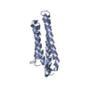
| ||||||||
| 2 | 
| ||||||||
| 3 | 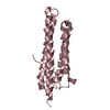
| ||||||||
| 4 | 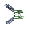
| ||||||||
| 5 | 
| ||||||||
| 6 | 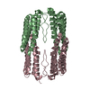
| ||||||||
| 7 | 
| ||||||||
| Unit cell |
|
-Components
| #1: Protein | Mass: 17949.697 Da / Num. of mol.: 3 Source method: isolated from a genetically manipulated source Source: (gene. exp.) Homo sapiens (human) / Gene: FAK / Plasmid: pGEX-6P2 / Production host:  #2: Water | ChemComp-HOH / |  Mass: 18.015 Da / Num. of mol.: 6 / Source method: isolated from a natural source / Formula: H2O Mass: 18.015 Da / Num. of mol.: 6 / Source method: isolated from a natural source / Formula: H2O |
|---|
-Experimental details
-Experiment
| Experiment | Method: X-RAY DIFFRACTION / Number of used crystals: 1 |
|---|
- Sample preparation
| Crystal | Density Matthews: 4.42 Å3/Da / Density % sol: 71.95 % | |||||||||||||||||||||||||||||||||||||||||||||||||||||||||||||||
|---|---|---|---|---|---|---|---|---|---|---|---|---|---|---|---|---|---|---|---|---|---|---|---|---|---|---|---|---|---|---|---|---|---|---|---|---|---|---|---|---|---|---|---|---|---|---|---|---|---|---|---|---|---|---|---|---|---|---|---|---|---|---|---|---|
| Crystal grow | Temperature: 296 K / Method: vapor diffusion, hanging drop / pH: 7 Details: Hepes, NaCl, glycerol, pH 7.0, VAPOR DIFFUSION, HANGING DROP, temperature 296K | |||||||||||||||||||||||||||||||||||||||||||||||||||||||||||||||
| Crystal grow | *PLUS pH: 8 | |||||||||||||||||||||||||||||||||||||||||||||||||||||||||||||||
| Components of the solutions | *PLUS
|
-Data collection
| Diffraction | Mean temperature: 100 K |
|---|---|
| Diffraction source | Source: SYNCHROTRON / Site: ESRF  / Beamline: ID14-3 / Wavelength: 0.931 Å / Beamline: ID14-3 / Wavelength: 0.931 Å |
| Detector | Type: ADSC QUANTUM 4 / Detector: CCD / Date: Sep 2, 2000 |
| Radiation | Protocol: SINGLE WAVELENGTH / Monochromatic (M) / Laue (L): M / Scattering type: x-ray |
| Radiation wavelength | Wavelength: 0.931 Å / Relative weight: 1 |
| Reflection | Resolution: 2.9→31.3 Å / Num. obs: 20719 / % possible obs: 96.9 % / Observed criterion σ(F): 0.9 / Observed criterion σ(I): 0.9 / Redundancy: 2.4 % / Biso Wilson estimate: 90 Å2 / Rsym value: 0.069 / Net I/σ(I): 7.4 |
| Reflection shell | Resolution: 2.9→3 Å / Redundancy: 2.3 % / Mean I/σ(I) obs: 1.2 / Num. unique all: 4413 / Rsym value: 0.626 / % possible all: 94.9 |
| Reflection | *PLUS Num. measured all: 48770 / Rmerge(I) obs: 0.069 |
| Reflection shell | *PLUS % possible obs: 94.9 % / Rmerge(I) obs: 0.626 |
- Processing
| Software |
| |||||||||||||||||||||||||
|---|---|---|---|---|---|---|---|---|---|---|---|---|---|---|---|---|---|---|---|---|---|---|---|---|---|---|
| Refinement | Method to determine structure: MIRAS / Resolution: 2.9→31.3 Å / Isotropic thermal model: isotropic / σ(F): 0 / σ(I): 0 / Stereochemistry target values: CNS
| |||||||||||||||||||||||||
| Displacement parameters | Biso mean: 76.8 Å2
| |||||||||||||||||||||||||
| Refinement step | Cycle: LAST / Resolution: 2.9→31.3 Å
| |||||||||||||||||||||||||
| Refine LS restraints |
| |||||||||||||||||||||||||
| LS refinement shell | Resolution: 2.9→2.95 Å
| |||||||||||||||||||||||||
| Xplor file |
| |||||||||||||||||||||||||
| Refinement | *PLUS % reflection Rfree: 7 % / Rfactor obs: 0.245 / Rfactor Rfree: 0.285 / Rfactor Rwork: 0.245 | |||||||||||||||||||||||||
| Solvent computation | *PLUS | |||||||||||||||||||||||||
| Displacement parameters | *PLUS | |||||||||||||||||||||||||
| LS refinement shell | *PLUS Rfactor Rfree: 0.511 / Rfactor Rwork: 0.503 / Rfactor obs: 0.503 |
