 Movie
Movie Controller


+ Open data

- Basic information
| Entry | Database: PDB / ID: 7kq5 | ||||||||||||
|---|---|---|---|---|---|---|---|---|---|---|---|---|---|
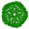
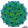
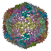
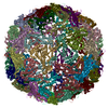
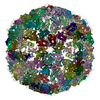
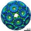
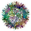
| Title | Cryo-EM structure of a thermostable encapsulin from T. maritima | ||||||||||||
 Components Components | Maritimacin | ||||||||||||
 Keywords Keywords | VIRUS LIKE PARTICLE / Encapsulin / HK97 fold / flavin-binding / icosahedral | ||||||||||||
| Function / homology |  Function and homology information Function and homology informationencapsulin nanocompartment / Hydrolases; Acting on peptide bonds (peptidases) / iron ion transport / peptidase activity / proteolysis Similarity search - Function | ||||||||||||
| Biological species |   Thermotoga maritima (bacteria) Thermotoga maritima (bacteria) | ||||||||||||
| Method | ELECTRON MICROSCOPY / single particle reconstruction / cryo EM / Resolution: 2 Å | ||||||||||||
 Authors Authors | Wiryaman, T.I. / Toor, N. | ||||||||||||
| Funding support |  United States, 3items United States, 3items
| ||||||||||||
 Citation Citation | Journal: IUCrJ / Year: 2021 Title: Cryo-EM structure of a thermostable bacterial nanocompartment. Authors: Timothy Wiryaman / Navtej Toor / Abstract: Protein nanocompartments are widespread in bacteria and archaea, but their functions are not yet well understood. Here, the cryo-EM structure of a nanocompartment from the thermophilic bacterium is ...Protein nanocompartments are widespread in bacteria and archaea, but their functions are not yet well understood. Here, the cryo-EM structure of a nanocompartment from the thermophilic bacterium is reported at 2.0 Å resolution. The high resolution of this structure shows that interactions in the E-loop domain may be important for the thermostability of the nanocompartment assembly. Also, the channels at the fivefold axis, threefold axis and dimer interface are assessed for their ability to transport iron. Finally, an unexpected flavin ligand was identified on the exterior of the shell, indicating that this nanocompartment may also play a direct role in iron metabolism. | ||||||||||||
| History |
|
- Structure visualization
| Movie |
Movie viewer |
|---|---|
| Structure viewer | Molecule: MolmilJmol/JSmol |
- Downloads & links
-Download
| PDBx/mmCIF format | 7kq5.cif.gz | 109.7 KB | Display | PDBx/mmCIF format |
|---|---|---|---|---|
| PDB format | pdb7kq5.ent.gz | 83.7 KB | Display | PDB format |
| PDBx/mmJSON format | 7kq5.json.gz | Tree view | PDBx/mmJSON format | |
| Others |  Other downloads Other downloads |
-Validation report
| Arichive directory | https://data.pdbj.org/pub/pdb/validation_reports/kq/7kq5ftp://data.pdbj.org/pub/pdb/validation_reports/kq/7kq5 | HTTPS FTP |
|---|
-Related structure data
| Related structure data |  22992MC M: map data used to model this data C: citing same article ( |
|---|---|
| Similar structure data | |
| EM raw data | EMPIAR-10674 (Title: Cryo-EM structure of a thermostable encapsulin from T. maritima Data size: 1.6 TB Data #1: Unaligned multiframe movies of T. maritima encapsulin [micrographs - multiframe]) |


-Links
 PDBj
PDBj
- Assembly
| Deposited unit | 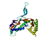
|
|---|---|
| 1 | x 60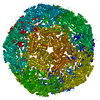
|
| 2 |
|
| 3 | x 5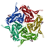
|
| 4 | x 6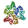
|
| 5 | 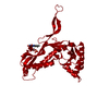
|
| Symmetry | Point symmetry: (Schoenflies symbol: I (icosahedral)) |
-Components
| #1: Protein | Mass: 30516.787 Da / Num. of mol.: 1 Source method: isolated from a genetically manipulated source Source: (gene. exp.) Thermotoga maritima (strain ATCC 43589 / MSB8 / DSM 3109 / JCM 10099) (bacteria)Strain: ATCC 43589 / MSB8 / DSM 3109 / JCM 10099 / Gene: TM_0785 / Plasmid: pET-11a / Production host: References: UniProt: Q9WZP2, Hydrolases; Acting on peptide bonds (peptidases) |
|---|---|
| #2: Chemical | ChemComp-FMN /   Mass: 456.344 Da / Num. of mol.: 1 / Source method: obtained synthetically / Formula: C17H21N4O9P Mass: 456.344 Da / Num. of mol.: 1 / Source method: obtained synthetically / Formula: C17H21N4O9P |
| #3: Water | ChemComp-HOH /  Mass: 18.015 Da / Num. of mol.: 148 / Source method: isolated from a natural source / Formula: H2O Mass: 18.015 Da / Num. of mol.: 148 / Source method: isolated from a natural source / Formula: H2O |
| Has ligand of interest | N |
-Experimental details
-Experiment
| Experiment | Method: ELECTRON MICROSCOPY |
|---|---|
| EM experiment | Aggregation state: PARTICLE / 3D reconstruction method: single particle reconstruction |
- Sample preparation
| Component | Name: Encapsulin / Type: COMPLEX / Entity ID: #1 / Source: RECOMBINANT | |||||||||||||||
|---|---|---|---|---|---|---|---|---|---|---|---|---|---|---|---|---|
| Molecular weight | Value: 1.82868 MDa / Experimental value: NO | |||||||||||||||
| Source (natural) | Organism: Thermotoga maritima MSB8 (bacteria) | |||||||||||||||
| Source (recombinant) | Organism: | |||||||||||||||
| Buffer solution | pH: 8 | |||||||||||||||
| Buffer component |
| |||||||||||||||
| Specimen | Conc.: 2 mg/ml / Embedding applied: NO / Shadowing applied: NO / Staining applied: NO / Vitrification applied: YES | |||||||||||||||
| Specimen support | Details: Plasma cleaned in Gatan Solarus system / Grid material: COPPER / Grid mesh size: 300 divisions/in. / Grid type: Quantifoil R1.2/1.3 | |||||||||||||||
| Vitrification | Instrument: LEICA EM GP / Cryogen name: PROPANE / Humidity: 90 % / Chamber temperature: 277 K Details: 3 ul sample applied to the carbon side of the grid and blotted for 4s before plunging. |
- Electron microscopy imaging
| Experimental equipment |  Model: Titan Krios / Image courtesy: FEI Company |
|---|---|
| Microscopy | Model: FEI TITAN KRIOS |
| Electron gun | Electron source:  FIELD EMISSION GUN / Accelerating voltage: 300 kV / Illumination mode: SPOT SCAN FIELD EMISSION GUN / Accelerating voltage: 300 kV / Illumination mode: SPOT SCAN |
| Electron lens | Mode: BRIGHT FIELD / Nominal magnification: 81000 X / Nominal defocus max: 2500 nm / Nominal defocus min: 500 nm / Cs: 2.7 mm |
| Specimen holder | Specimen holder model: FEI TITAN KRIOS AUTOGRID HOLDER |
| Image recording | Average exposure time: 2.5 sec. / Electron dose: 33.5 e/Å2 / Film or detector model: GATAN K3 (6k x 4k) / Num. of real images: 2772 |
| Image scans | Width: 11520 / Height: 8184 |
- Processing
| Software | Name: PHENIX / Version: 1.18.2_3874: / Classification: refinement | |||||||||||||||||||||||||||||||||||||||||||||
|---|---|---|---|---|---|---|---|---|---|---|---|---|---|---|---|---|---|---|---|---|---|---|---|---|---|---|---|---|---|---|---|---|---|---|---|---|---|---|---|---|---|---|---|---|---|---|
| EM software |
| |||||||||||||||||||||||||||||||||||||||||||||
| CTF correction | Type: NONE | |||||||||||||||||||||||||||||||||||||||||||||
| Particle selection | Num. of particles selected: 204251 / Details: Picked with trained cryolo model | |||||||||||||||||||||||||||||||||||||||||||||
| Symmetry | Point symmetry: I (icosahedral) | |||||||||||||||||||||||||||||||||||||||||||||
| 3D reconstruction | Resolution: 2 Å / Resolution method: FSC 0.143 CUT-OFF / Num. of particles: 185459 / Num. of class averages: 1 / Symmetry type: POINT | |||||||||||||||||||||||||||||||||||||||||||||
| Atomic model building | Protocol: OTHER Details: Map was cropped to a 342 box size and density modified with PHENIX ResolveCryoEM | |||||||||||||||||||||||||||||||||||||||||||||
| Atomic model building | PDB-ID: 3DKT Pdb chain-ID: A / Accession code: 3DKT / Pdb chain residue range: 4-269 / Source name: PDB / Type: experimental model | |||||||||||||||||||||||||||||||||||||||||||||
| Refine LS restraints |
|

