Mass: 18.015 Da / Num. of mol.: 12 / Source method: isolated from a natural source / Formula: H2O
Compound details
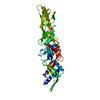
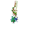
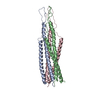
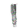
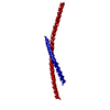
THIS STRUCTURE IS A CHIMERA. IT STARTS AT THE N-TERMINUS WITH 31 RESIDUES FROM YEAST GCN4 FOLLOWED ...THIS STRUCTURE IS A CHIMERA. IT STARTS AT THE N-TERMINUS WITH 31 RESIDUES FROM YEAST GCN4 FOLLOWED BY 50 RESIDUES FROM GP41 AND 42 ADDITIONAL RESIDUES FROM GP41, FOR A A TOTAL OF 123 RESIDUES AS SHOWN ON SEQRES. 115 OF THESE RESIDUES WERE LOCATED AND ARE PRESENTED IN THIS ENTRY. DBREF RECORDS BELOW CAN BE USED TO ASSOCIATE THE RESIDUES PRESENT IN THIS ENTRY WITH THE CORRESPONDING SWISSPROT ENTRY.
-
Experimental details
-
Experiment
Experiment
Method: X-RAY DIFFRACTION / Number of used crystals: 1
-
Sample preparation
Crystal
Density Matthews: 4.4 Å3/Da / Density % sol: 72 %
Crystal grow
pH: 8 Details: 20 MM HEPES PH 8.0 1.1 M AMMONIUM SULFATE 12% ETHYLENE GLYCOL SPACE GROUP R32 IN HEXAGONAL SETTING
In the structure databanks used in Yorodumi, some data are registered as the other names, "COVID-19 virus" and "2019-nCoV". Here are the details of the virus and the list of structure data.
Jan 31, 2019. EMDB accession codes are about to change! (news from PDBe EMDB page)
EMDB accession codes are about to change! (news from PDBe EMDB page)
The allocation of 4 digits for EMDB accession codes will soon come to an end. Whilst these codes will remain in use, new EMDB accession codes will include an additional digit and will expand incrementally as the available range of codes is exhausted. The current 4-digit format prefixed with “EMD-” (i.e. EMD-XXXX) will advance to a 5-digit format (i.e. EMD-XXXXX), and so on. It is currently estimated that the 4-digit codes will be depleted around Spring 2019, at which point the 5-digit format will come into force.
The EM Navigator/Yorodumi systems omit the EMD- prefix.
Related info.:Q: What is EMD? / ID/Accession-code notation in Yorodumi/EM Navigator
Yorodumi is a browser for structure data from EMDB, PDB, SASBDB, etc.
This page is also the successor to EM Navigator detail page, and also detail information page/front-end page for Omokage search.
The word "yorodu" (or yorozu) is an old Japanese word meaning "ten thousand". "mi" (miru) is to see.
Related info.:EMDB / PDB / SASBDB / Comparison of 3 databanks / Yorodumi Search / Aug 31, 2016. New EM Navigator & Yorodumi / Yorodumi Papers / Jmol/JSmol / Function and homology information / Changes in new EM Navigator and Yorodumi
 Movie
Movie Controller


 Open data
 Basic information
 Components
 Keywords
 Function and homology information

 Human immunodeficiency virus 1
 X-RAY DIFFRACTION /
  Authors
 Citation
 Structure visualization
 Downloads & links
 Other downloads


 PDBj
PDBj


 Assembly
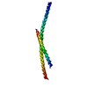

 Mass: 18.015 Da / Num. of mol.: 12 / Source method: isolated from a natural source / Formula: H2O
 Sample preparation
 Processing