 Movie
Movie Controller

[English] 日本語
 Yorodumi
- PDB-1kql: Crystal structure of the C-terminal region of striated muscle alp... -
+ Open data

- Basic information
| Entry | Database: PDB / ID: 1kql | ||||||
|---|---|---|---|---|---|---|---|
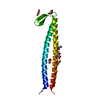
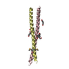
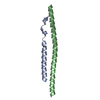
| Title | Crystal structure of the C-terminal region of striated muscle alpha-tropomyosin at 2.7 angstrom resolution | ||||||
 Components Components | Fusion Protein of and striated muscle alpha-tropomyosin and the GCN4 leucine zipper | ||||||
 Keywords Keywords | CONTRACTILE PROTEIN / thin filament / tropomyosin / muscle regulation / coiled coil / four-helix bundle | ||||||
| Function / homology |  Function and homology information Function and homology informationStriated Muscle Contraction / Smooth Muscle Contraction / positive regulation of heart rate by epinephrine / FCERI mediated MAPK activation / protein localization to nuclear periphery / Activation of the AP-1 family of transcription factors / negative regulation of ribosomal protein gene transcription by RNA polymerase II / positive regulation of cellular response to amino acid starvation / response to amino acid starvation / mediator complex binding ...Striated Muscle Contraction / Smooth Muscle Contraction / positive regulation of heart rate by epinephrine / FCERI mediated MAPK activation / protein localization to nuclear periphery / Activation of the AP-1 family of transcription factors / negative regulation of ribosomal protein gene transcription by RNA polymerase II / positive regulation of cellular response to amino acid starvation / response to amino acid starvation / mediator complex binding / bleb / ruffle organization / Oxidative Stress Induced Senescence / actin filament capping / muscle filament sliding / sarcomere organization / ventricular cardiac muscle tissue morphogenesis / negative regulation of vascular associated smooth muscle cell migration / amino acid biosynthetic process / TFIID-class transcription factor complex binding / myofibril / negative regulation of vascular associated smooth muscle cell proliferation / positive regulation of RNA polymerase II transcription preinitiation complex assembly / positive regulation of transcription initiation by RNA polymerase II / cytoskeletal protein binding / positive regulation of stress fiber assembly / cardiac muscle contraction / stress fiber / cellular response to nutrient levels / positive regulation of cell adhesion / muscle contraction / negative regulation of cell migration / cellular response to amino acid starvation / actin filament organization / actin filament / wound healing / cellular response to reactive oxygen species / RNA polymerase II transcription regulator complex / ruffle membrane / disordered domain specific binding / actin filament binding / regulation of cell shape / actin cytoskeleton / actin binding / DNA-binding transcription activator activity, RNA polymerase II-specific / transcription regulator complex / sequence-specific DNA binding / in utero embryonic development / RNA polymerase II-specific DNA-binding transcription factor binding / DNA-binding transcription factor activity, RNA polymerase II-specific / intracellular signal transduction / RNA polymerase II cis-regulatory region sequence-specific DNA binding / DNA-binding transcription factor activity / protein heterodimerization activity / chromatin binding / negative regulation of transcription by RNA polymerase II / protein homodimerization activity / positive regulation of transcription by RNA polymerase II / protein-containing complex / identical protein binding / nucleus / cytoplasm / cytosol Similarity search - Function | ||||||
| Biological species |   | ||||||
| Method |  X-RAY DIFFRACTION / SYNCHROTRON / Molecular replacement using a specially built model / Resolution: 2.7 Å X-RAY DIFFRACTION / SYNCHROTRON / Molecular replacement using a specially built model / Resolution: 2.7 Å | ||||||
 Authors Authors | Li, Y. / Mui, S. / Brown, J.H. / Strand, J. / Reshetnikova, L. / Tobacman, L.S. / Cohen, C. | ||||||
 Citation Citation | Journal: Proc.Natl.Acad.Sci.USA / Year: 2002 Title: The crystal structure of the C-terminal fragment of striated-muscle alpha-tropomyosin reveals a key troponin T recognition site. Authors: Li, Y. / Mui, S. / Brown, J.H. / Strand, J. / Reshetnikova, L. / Tobacman, L.S. / Cohen, C. | ||||||
| History |
|
- Structure visualization
| Structure viewer | Molecule: MolmilJmol/JSmol |
|---|
- Downloads & links
-Download
| PDBx/mmCIF format | 1kql.cif.gz | 33.4 KB | Display | PDBx/mmCIF format |
|---|---|---|---|---|
| PDB format | pdb1kql.ent.gz | 24.3 KB | Display | PDB format |
| PDBx/mmJSON format | 1kql.json.gz | Tree view | PDBx/mmJSON format | |
| Others |  Other downloads Other downloads |
-Validation report
| Arichive directory | https://data.pdbj.org/pub/pdb/validation_reports/kq/1kqlftp://data.pdbj.org/pub/pdb/validation_reports/kq/1kql | HTTPS FTP |
|---|
-Related structure data
| Similar structure data |
|---|


-Links
 PDBj
PDBj

- Assembly
| Deposited unit | 
| ||||||||
|---|---|---|---|---|---|---|---|---|---|
| 1 |
| ||||||||
| 2 | 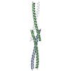
| ||||||||
| Unit cell |
| ||||||||
| Components on special symmetry positions |
|
-Components
| #1: Protein | Mass: 6603.480 Da / Num. of mol.: 2 Source method: isolated from a genetically manipulated source Details: N-terminal methionine followed by sequence database residues 255-278 of GCN4 leucine zipper and then C-terminal sequence database residues 254-284 of rat striated muscle alpha-tropomyosin Source: (gene. exp.) Genus: Saccharomyces, Rattus / Species: , / Strain: , / Plasmid: pET3d / Gene (production host): GCN4, ARG9 / Production host:  References: UniProt: P03069, GenBank: 207514, UniProt: P04692*PLUS #2: Water | ChemComp-HOH / |  Mass: 18.015 Da / Num. of mol.: 27 / Source method: isolated from a natural source / Formula: H2O Mass: 18.015 Da / Num. of mol.: 27 / Source method: isolated from a natural source / Formula: H2O |
|---|
-Experimental details
-Experiment
| Experiment | Method: X-RAY DIFFRACTION / Number of used crystals: 1 |
|---|
- Sample preparation
| Crystal | Density Matthews: 4.12 Å3/Da / Density % sol: 70 % | |||||||||||||||||||||||||||||||||||||||||||||||||||||||||||||||||||||||||||||||||||||||||||
|---|---|---|---|---|---|---|---|---|---|---|---|---|---|---|---|---|---|---|---|---|---|---|---|---|---|---|---|---|---|---|---|---|---|---|---|---|---|---|---|---|---|---|---|---|---|---|---|---|---|---|---|---|---|---|---|---|---|---|---|---|---|---|---|---|---|---|---|---|---|---|---|---|---|---|---|---|---|---|---|---|---|---|---|---|---|---|---|---|---|---|---|---|
| Crystal grow | Temperature: 295 K / Method: vapor diffusion, sitting drop / pH: 8.3 Details: PEG 550 monomethylether, glycerol, sodium chloride, magnesium acetate, bicine, tris buffer, dithiothreitol, pH 8.3, VAPOR DIFFUSION, SITTING DROP, temperature 295K | |||||||||||||||||||||||||||||||||||||||||||||||||||||||||||||||||||||||||||||||||||||||||||
| Crystal grow | *PLUS Temperature: 22 ℃ / pH: 7.5 / Method: vapor diffusion | |||||||||||||||||||||||||||||||||||||||||||||||||||||||||||||||||||||||||||||||||||||||||||
| Components of the solutions | *PLUS
|
-Data collection
| Diffraction | Mean temperature: 95 K |
|---|---|
| Diffraction source | Source: SYNCHROTRON / Site: CHESS  / Beamline: A1 / Wavelength: 0.909 Å / Beamline: A1 / Wavelength: 0.909 Å |
| Detector | Type: ADSC QUANTUM 4 / Detector: CCD / Date: Apr 20, 2001 / Details: mirrors |
| Radiation | Monochromator: GRAPHITE / Protocol: SINGLE WAVELENGTH / Monochromatic (M) / Laue (L): M / Scattering type: x-ray |
| Radiation wavelength | Wavelength: 0.909 Å / Relative weight: 1 |
| Reflection | Resolution: 2.6→50 Å / Num. all: 7229 / Num. obs: 7221 / % possible obs: 99.9 % / Observed criterion σ(F): 2 / Observed criterion σ(I): 2 / Redundancy: 12 % / Biso Wilson estimate: 82.3 Å2 / Rmerge(I) obs: 0.052 / Net I/σ(I): 34.9 |
| Reflection shell | Resolution: 2.6→2.69 Å / Redundancy: 8.2 % / Rmerge(I) obs: 0.217 / Mean I/σ(I) obs: 9.14 / Num. unique all: 708 / % possible all: 99.9 |
| Reflection | *PLUS Lowest resolution: 9999 Å / Num. measured all: 86920 / Rmerge(I) obs: 0.052 |
| Reflection shell | *PLUS % possible obs: 99.9 % / Rmerge(I) obs: 0.217 |
- Processing
| Software |
| |||||||||||||||||||||||||||||||||||||||||||||||||
|---|---|---|---|---|---|---|---|---|---|---|---|---|---|---|---|---|---|---|---|---|---|---|---|---|---|---|---|---|---|---|---|---|---|---|---|---|---|---|---|---|---|---|---|---|---|---|---|---|---|---|
| Refinement | Method to determine structure: Molecular replacement using a specially built model Starting model: a specially built 56-residue two-stranded coiled-coil that contains a 24 residue fragment from the high resolution structure of the GCN4 leucine zipper Resolution: 2.7→44.91 Å / Isotropic thermal model: RESTRAINED / Cross valid method: THROUGHOUT / σ(F): 0 / σ(I): 0 / Stereochemistry target values: Engh & Huber
| |||||||||||||||||||||||||||||||||||||||||||||||||
| Displacement parameters | Biso mean: 93.4 Å2
| |||||||||||||||||||||||||||||||||||||||||||||||||
| Refine analyze |
| |||||||||||||||||||||||||||||||||||||||||||||||||
| Refinement step | Cycle: LAST / Resolution: 2.7→44.91 Å
| |||||||||||||||||||||||||||||||||||||||||||||||||
| Refine LS restraints |
| |||||||||||||||||||||||||||||||||||||||||||||||||
| LS refinement shell | Refine-ID: X-RAY DIFFRACTION / Total num. of bins used: 10
| |||||||||||||||||||||||||||||||||||||||||||||||||
| Refinement | *PLUS Highest resolution: 2.7 Å / Lowest resolution: 9999 Å / Num. reflection obs: 6498 / % reflection Rfree: 8 % / Rfactor all: 0.2551 / Rfactor obs: 0.251 / Rfactor Rfree: 0.289 / Rfactor Rwork: 0.251 | |||||||||||||||||||||||||||||||||||||||||||||||||
| Solvent computation | *PLUS | |||||||||||||||||||||||||||||||||||||||||||||||||
| Displacement parameters | *PLUS | |||||||||||||||||||||||||||||||||||||||||||||||||
| Refine LS restraints | *PLUS
| |||||||||||||||||||||||||||||||||||||||||||||||||
| LS refinement shell | *PLUS Highest resolution: 2.6 Å / Lowest resolution: 2.69 Å / Rfactor Rfree: 0.3353 / Rfactor Rwork: 0.3295 / Rfactor obs: 0.3295 |
