 Movie
Movie Controller


+ Open data

- Basic information
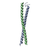
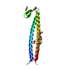
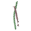
| Entry | Database: PDB / ID: 5vpa | |||||||||
|---|---|---|---|---|---|---|---|---|---|---|
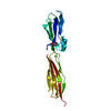
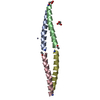
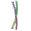
| Title | Transcription factor FosB/JunD bZIP domain | |||||||||
 Components Components |
| |||||||||
 Keywords Keywords | TRANSCRIPTION / activator protein-1 / basic leucine zipper / bZIP / fos / jun / transcription factor / DNA-binding protein / redox switch / coiled-coil | |||||||||
| Function / homology |  Function and homology information Function and homology informationresponse to forskolin / transcription factor AP-1 complex / positive regulation of peroxisome proliferator activated receptor signaling pathway / NGF-stimulated transcription / positive regulation of macrophage activation / cellular response to fatty acid / osteoblast development / response to steroid hormone / response to morphine / response to corticosterone ...response to forskolin / transcription factor AP-1 complex / positive regulation of peroxisome proliferator activated receptor signaling pathway / NGF-stimulated transcription / positive regulation of macrophage activation / cellular response to fatty acid / osteoblast development / response to steroid hormone / response to morphine / response to corticosterone / positive regulation of osteoblast differentiation / response to light stimulus / response to cAMP / response to mechanical stimulus / cellular response to hormone stimulus / behavioral response to cocaine / transcription repressor complex / response to progesterone / cellular response to calcium ion / response to amphetamine / nuclear receptor binding / transcription coregulator binding / response to nicotine / female pregnancy / circadian rhythm / protein-DNA complex / RNA polymerase II transcription regulator complex / response to peptide hormone / sequence-specific double-stranded DNA binding / regulation of cell population proliferation / transcription by RNA polymerase II / DNA-binding transcription activator activity, RNA polymerase II-specific / transcription regulator complex / response to lipopolysaccharide / cellular response to hypoxia / Estrogen-dependent gene expression / response to ethanol / DNA-binding transcription factor activity, RNA polymerase II-specific / transcription coactivator activity / regulation of cell cycle / transcription cis-regulatory region binding / response to xenobiotic stimulus / RNA polymerase II cis-regulatory region sequence-specific DNA binding / regulation of transcription by RNA polymerase II / chromatin / enzyme binding / negative regulation of transcription by RNA polymerase II / positive regulation of transcription by RNA polymerase II / DNA binding / nucleoplasm / nucleus / cytosol Similarity search - Function | |||||||||
| Biological species |  Homo sapiens (human) Homo sapiens (human) | |||||||||
| Method |  X-RAY DIFFRACTION / SYNCHROTRON / MOLECULAR REPLACEMENT / Resolution: 2.83 Å X-RAY DIFFRACTION / SYNCHROTRON / MOLECULAR REPLACEMENT / Resolution: 2.83 Å | |||||||||
 Authors Authors | Yin, Z. / Machius, M. / Rudenko, G. | |||||||||
| Funding support |  United States, 2items United States, 2items
| |||||||||
 Citation Citation | Journal: Nucleic Acids Res. / Year: 2017 Title: Activator Protein-1: redox switch controlling structure and DNA-binding. Authors: Yin, Z. / Machius, M. / Nestler, E.J. / Rudenko, G. | |||||||||
| History |
|
- Structure visualization
| Structure viewer | Molecule: MolmilJmol/JSmol |
|---|
- Downloads & links
-Download
| PDBx/mmCIF format | 5vpa.cif.gz | 80.7 KB | Display | PDBx/mmCIF format |
|---|---|---|---|---|
| PDB format | pdb5vpa.ent.gz | 61.7 KB | Display | PDB format |
| PDBx/mmJSON format | 5vpa.json.gz | Tree view | PDBx/mmJSON format | |
| Others |  Other downloads Other downloads |
-Validation report
| Arichive directory | https://data.pdbj.org/pub/pdb/validation_reports/vp/5vpaftp://data.pdbj.org/pub/pdb/validation_reports/vp/5vpa | HTTPS FTP |
|---|
-Related structure data
| Related structure data |  5vpbC  5vpcC  5vpdC  5vpeC  5vpfC 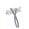 1fosS C: citing same article ( S: Starting model for refinement |
|---|---|
| Similar structure data |


-Links
 PDBj
PDBj


- Assembly
| Deposited unit | 
| ||||||||
|---|---|---|---|---|---|---|---|---|---|
| 1 |
| ||||||||
| Unit cell |
|
-Components
| #1: Protein | Mass: 8267.290 Da / Num. of mol.: 1 / Fragment: unp residues 153-219 Source method: isolated from a genetically manipulated source Source: (gene. exp.) Homo sapiens (human) / Gene: FOSB, G0S3 / Plasmid: pET21-NESG / Production host:  |
|---|---|
| #2: Protein | Mass: 7989.425 Da / Num. of mol.: 1 / Fragment: unp residues 266-332 Source method: isolated from a genetically manipulated source Source: (gene. exp.) Homo sapiens (human) / Gene: JUND / Plasmid: pET21a / Production host: |
| #3: Chemical | ChemComp-NA /   Mass: 22.990 Da / Num. of mol.: 1 / Source method: obtained synthetically / Formula: Na Mass: 22.990 Da / Num. of mol.: 1 / Source method: obtained synthetically / Formula: Na |
| #4: Chemical | ChemComp-CL /   Mass: 35.453 Da / Num. of mol.: 1 / Source method: obtained synthetically / Formula: Cl Mass: 35.453 Da / Num. of mol.: 1 / Source method: obtained synthetically / Formula: Cl |
| #5: Water | ChemComp-HOH /  Mass: 18.015 Da / Num. of mol.: 5 / Source method: isolated from a natural source / Formula: H2O Mass: 18.015 Da / Num. of mol.: 5 / Source method: isolated from a natural source / Formula: H2O |
| Has protein modification | Y |
-Experimental details
-Experiment
| Experiment | Method: X-RAY DIFFRACTION / Number of used crystals: 1 |
|---|
- Sample preparation
| Crystal | Density Matthews: 3.79 Å3/Da / Density % sol: 67.52 % / Description: thin plates |
|---|---|
| Crystal grow | Temperature: 293 K / Method: vapor diffusion, sitting drop / pH: 8.5 Details: 3 M 1,6-Hexanediol, 50 mM Tris pH 8.5 and 5 mM MgCl2 |
-Data collection
| Diffraction | Mean temperature: 100 K | |||||||||||||||||||||||||||||||||||||||||||||||||||||||||||||||||||||||||||||||||||||||||||||||||||||||||||||||||||||||||||||||||||||||||||||||||||||||||||||||||||||||||||||||||||||||||||||
|---|---|---|---|---|---|---|---|---|---|---|---|---|---|---|---|---|---|---|---|---|---|---|---|---|---|---|---|---|---|---|---|---|---|---|---|---|---|---|---|---|---|---|---|---|---|---|---|---|---|---|---|---|---|---|---|---|---|---|---|---|---|---|---|---|---|---|---|---|---|---|---|---|---|---|---|---|---|---|---|---|---|---|---|---|---|---|---|---|---|---|---|---|---|---|---|---|---|---|---|---|---|---|---|---|---|---|---|---|---|---|---|---|---|---|---|---|---|---|---|---|---|---|---|---|---|---|---|---|---|---|---|---|---|---|---|---|---|---|---|---|---|---|---|---|---|---|---|---|---|---|---|---|---|---|---|---|---|---|---|---|---|---|---|---|---|---|---|---|---|---|---|---|---|---|---|---|---|---|---|---|---|---|---|---|---|---|---|---|---|---|
| Diffraction source | Source: SYNCHROTRON / Site: APS / Beamline: 17-ID / Wavelength: 0.999 Å | |||||||||||||||||||||||||||||||||||||||||||||||||||||||||||||||||||||||||||||||||||||||||||||||||||||||||||||||||||||||||||||||||||||||||||||||||||||||||||||||||||||||||||||||||||||||||||||
| Detector | Type: DECTRIS PILATUS3 S 6M / Detector: PIXEL / Date: Nov 13, 2016 | |||||||||||||||||||||||||||||||||||||||||||||||||||||||||||||||||||||||||||||||||||||||||||||||||||||||||||||||||||||||||||||||||||||||||||||||||||||||||||||||||||||||||||||||||||||||||||||
| Radiation | Monochromator: Si(111) / Protocol: SINGLE WAVELENGTH / Monochromatic (M) / Laue (L): M / Scattering type: x-ray | |||||||||||||||||||||||||||||||||||||||||||||||||||||||||||||||||||||||||||||||||||||||||||||||||||||||||||||||||||||||||||||||||||||||||||||||||||||||||||||||||||||||||||||||||||||||||||||
| Radiation wavelength | Wavelength: 0.999 Å / Relative weight: 1 | |||||||||||||||||||||||||||||||||||||||||||||||||||||||||||||||||||||||||||||||||||||||||||||||||||||||||||||||||||||||||||||||||||||||||||||||||||||||||||||||||||||||||||||||||||||||||||||
| Reflection | Resolution: 2.83→34.204 Å / Num. obs: 6010 / % possible obs: 100 % / Redundancy: 9.5 % / Biso Wilson estimate: 32.12 Å2 / Rmerge(I) obs: 0.096 / Rpim(I) all: 0.033 / Rrim(I) all: 0.102 / Χ2: 0.976 / Net I/σ(I): 25 | |||||||||||||||||||||||||||||||||||||||||||||||||||||||||||||||||||||||||||||||||||||||||||||||||||||||||||||||||||||||||||||||||||||||||||||||||||||||||||||||||||||||||||||||||||||||||||||
| Reflection shell | Diffraction-ID: 1
|
- Processing
| Software |
| ||||||||||||||||||||||||||||||||||||||||||||||||||||||||||||||||||||||||||||||||||||||||||||||||||||
|---|---|---|---|---|---|---|---|---|---|---|---|---|---|---|---|---|---|---|---|---|---|---|---|---|---|---|---|---|---|---|---|---|---|---|---|---|---|---|---|---|---|---|---|---|---|---|---|---|---|---|---|---|---|---|---|---|---|---|---|---|---|---|---|---|---|---|---|---|---|---|---|---|---|---|---|---|---|---|---|---|---|---|---|---|---|---|---|---|---|---|---|---|---|---|---|---|---|---|---|---|---|
| Refinement | Method to determine structure: MOLECULAR REPLACEMENT Starting model: 1FOS Resolution: 2.83→34.204 Å / SU ML: 0.38 / Cross valid method: FREE R-VALUE / σ(F): 1.35 / Phase error: 31.57
| ||||||||||||||||||||||||||||||||||||||||||||||||||||||||||||||||||||||||||||||||||||||||||||||||||||
| Solvent computation | Shrinkage radii: 0.9 Å / VDW probe radii: 1.11 Å | ||||||||||||||||||||||||||||||||||||||||||||||||||||||||||||||||||||||||||||||||||||||||||||||||||||
| Displacement parameters | Biso max: 185.72 Å2 / Biso mean: 66.4615 Å2 / Biso min: 4.48 Å2 | ||||||||||||||||||||||||||||||||||||||||||||||||||||||||||||||||||||||||||||||||||||||||||||||||||||
| Refinement step | Cycle: final / Resolution: 2.83→34.204 Å
| ||||||||||||||||||||||||||||||||||||||||||||||||||||||||||||||||||||||||||||||||||||||||||||||||||||
| Refine LS restraints |
| ||||||||||||||||||||||||||||||||||||||||||||||||||||||||||||||||||||||||||||||||||||||||||||||||||||
| LS refinement shell | Refine-ID: X-RAY DIFFRACTION / Rfactor Rfree error: 0 / Total num. of bins used: 3
| ||||||||||||||||||||||||||||||||||||||||||||||||||||||||||||||||||||||||||||||||||||||||||||||||||||
| Refinement TLS params. | Method: refined / Refine-ID: X-RAY DIFFRACTION
| ||||||||||||||||||||||||||||||||||||||||||||||||||||||||||||||||||||||||||||||||||||||||||||||||||||
| Refinement TLS group |
|
