 Movie
Movie Controller


+ Open data

- Basic information
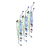
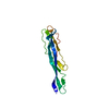
| Entry | Database: PDB / ID: 1ce9 | ||||||
|---|---|---|---|---|---|---|---|
| Title | HELIX CAPPING IN THE GCN4 LEUCINE ZIPPER | ||||||
 Components Components | PROTEIN (GCN4-PMSE) | ||||||
 Keywords Keywords | HELIX CAPPING / LEUCINE ZIPPER / HYDROGEN BONDING / THERMAL STABILITY / PROTEIN FOLDING | ||||||
| Function / homology |  Function and homology information Function and homology informationActivation of the AP-1 family of transcription factors / FCERI mediated MAPK activation / protein localization to nuclear periphery / negative regulation of ribosomal protein gene transcription by RNA polymerase II / Oxidative Stress Induced Senescence / positive regulation of cellular response to amino acid starvation / response to amino acid starvation / mediator complex binding / amino acid biosynthetic process / TFIID-class transcription factor complex binding ...Activation of the AP-1 family of transcription factors / FCERI mediated MAPK activation / protein localization to nuclear periphery / negative regulation of ribosomal protein gene transcription by RNA polymerase II / Oxidative Stress Induced Senescence / positive regulation of cellular response to amino acid starvation / response to amino acid starvation / mediator complex binding / amino acid biosynthetic process / TFIID-class transcription factor complex binding / positive regulation of transcription initiation by RNA polymerase II / positive regulation of RNA polymerase II transcription preinitiation complex assembly / cellular response to nutrient levels / cellular response to amino acid starvation / RNA polymerase II transcription regulator complex / transcription regulator complex / DNA-binding transcription activator activity, RNA polymerase II-specific / sequence-specific DNA binding / RNA polymerase II-specific DNA-binding transcription factor binding / DNA-binding transcription factor activity, RNA polymerase II-specific / intracellular signal transduction / RNA polymerase II cis-regulatory region sequence-specific DNA binding / DNA-binding transcription factor activity / chromatin binding / negative regulation of transcription by RNA polymerase II / positive regulation of transcription by RNA polymerase II / identical protein binding / nucleus Similarity search - Function | ||||||
| Method |  X-RAY DIFFRACTION / MOLECULAR REPLACEMENT / Resolution: 1.8 Å X-RAY DIFFRACTION / MOLECULAR REPLACEMENT / Resolution: 1.8 Å | ||||||
 Authors Authors | Lu, M. / Shu, W. / Ji, H. / Spek, E. / Wang, L.-Y. / Kallenbach, N.R. | ||||||
 Citation Citation | Journal: J.Mol.Biol. / Year: 1999 Title: Helix capping in the GCN4 leucine zipper. Authors: Lu, M. / Shu, W. / Ji, H. / Spek, E. / Wang, L. / Kallenbach, N.R. #1: Journal: Science / Year: 1991Title: X-Ray Structure of the GCN4 Leucine Zipper, a Two-Stranded, Parallel Coiled Coil Authors: O'Shea, E.K. / Klemm, J.D. / Kim, P.S. / Alber, T. | ||||||
| History |
|
- Structure visualization
| Structure viewer | Molecule: MolmilJmol/JSmol |
|---|
- Downloads & links
-Download
| PDBx/mmCIF format | 1ce9.cif.gz | 38.9 KB | Display | PDBx/mmCIF format |
|---|---|---|---|---|
| PDB format | pdb1ce9.ent.gz | 28.6 KB | Display | PDB format |
| PDBx/mmJSON format | 1ce9.json.gz | Tree view | PDBx/mmJSON format | |
| Others |  Other downloads Other downloads |
-Validation report
| Arichive directory | https://data.pdbj.org/pub/pdb/validation_reports/ce/1ce9ftp://data.pdbj.org/pub/pdb/validation_reports/ce/1ce9 | HTTPS FTP |
|---|
-Related structure data
| Related structure data | 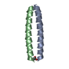 2ztaS S: Starting model for refinement |
|---|---|
| Similar structure data |


-Links
 PDBj
PDBj

- Assembly
| Deposited unit | 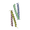
| ||||||||
|---|---|---|---|---|---|---|---|---|---|
| 1 |
| ||||||||
| 2 | 
| ||||||||
| 3 | 
| ||||||||
| Unit cell |
|
-Components
| #1: Protein/peptide | Mass: 4035.663 Da / Num. of mol.: 4 / Mutation: R2S,M3V,Q5E / Source method: obtained synthetically / References: UniProt: P03069 #2: Water | ChemComp-HOH / |  Mass: 18.015 Da / Num. of mol.: 150 / Source method: isolated from a natural source / Formula: H2O Mass: 18.015 Da / Num. of mol.: 150 / Source method: isolated from a natural source / Formula: H2O |
|---|
-Experimental details
-Experiment
| Experiment | Method: X-RAY DIFFRACTION / Number of used crystals: 1 |
|---|
- Sample preparation
| Crystal | Density Matthews: 2.5 Å3/Da / Density % sol: 50 % | |||||||||||||||||||||||||
|---|---|---|---|---|---|---|---|---|---|---|---|---|---|---|---|---|---|---|---|---|---|---|---|---|---|---|
| Crystal grow | pH: 4.6 / Details: SEE REFERENCE, pH 4.6 | |||||||||||||||||||||||||
| Crystal | *PLUS | |||||||||||||||||||||||||
| Crystal grow | *PLUS Method: vapor diffusion, hanging drop | |||||||||||||||||||||||||
| Components of the solutions | *PLUS
|
-Data collection
| Diffraction | Mean temperature: 298 K |
|---|---|
| Diffraction source | Source: ROTATING ANODE / Type: RIGAKU RU200 / Wavelength: 1.5418 |
| Detector | Type: RIGAKU RAXIS IV / Detector: IMAGE PLATE / Date: Mar 1, 1998 / Details: MIRRORS |
| Radiation | Monochromator: SUPPER DOUBLE MIRRORS / Protocol: SINGLE WAVELENGTH / Monochromatic (M) / Laue (L): M / Scattering type: x-ray |
| Radiation wavelength | Wavelength: 1.5418 Å / Relative weight: 1 |
| Reflection | Resolution: 1.8→30 Å / Num. obs: 11776 / % possible obs: 92.3 % / Observed criterion σ(I): 3 / Redundancy: 2 % / Rmerge(I) obs: 0.034 / Rsym value: 0.034 / Net I/σ(I): 13.6 |
| Reflection shell | Resolution: 1.8→1.86 Å / Redundancy: 1.9 % / Rmerge(I) obs: 0.059 / Mean I/σ(I) obs: 12 / Rsym value: 0.059 / % possible all: 88.7 |
| Reflection | *PLUS Num. measured all: 23475 |
- Processing
| Software |
| ||||||||||||||||||||||||||||||||||||||||||||||||||||||||||||
|---|---|---|---|---|---|---|---|---|---|---|---|---|---|---|---|---|---|---|---|---|---|---|---|---|---|---|---|---|---|---|---|---|---|---|---|---|---|---|---|---|---|---|---|---|---|---|---|---|---|---|---|---|---|---|---|---|---|---|---|---|---|
| Refinement | Method to determine structure: MOLECULAR REPLACEMENT Starting model: 2ZTA Resolution: 1.8→15 Å / Cross valid method: THROUGHOUT / σ(F): 0
| ||||||||||||||||||||||||||||||||||||||||||||||||||||||||||||
| Displacement parameters | Biso mean: 20.1 Å2 | ||||||||||||||||||||||||||||||||||||||||||||||||||||||||||||
| Refinement step | Cycle: LAST / Resolution: 1.8→15 Å
| ||||||||||||||||||||||||||||||||||||||||||||||||||||||||||||
| Refine LS restraints |
| ||||||||||||||||||||||||||||||||||||||||||||||||||||||||||||
| Software | *PLUS Name: X-PLOR / Version: 3.851 / Classification: refinement | ||||||||||||||||||||||||||||||||||||||||||||||||||||||||||||
| Refinement | *PLUS Rfactor obs: 0.214 | ||||||||||||||||||||||||||||||||||||||||||||||||||||||||||||
| Solvent computation | *PLUS | ||||||||||||||||||||||||||||||||||||||||||||||||||||||||||||
| Displacement parameters | *PLUS | ||||||||||||||||||||||||||||||||||||||||||||||||||||||||||||
| Refine LS restraints | *PLUS
|
