 Movie
Movie Controller


+ Open data

- Basic information
| Entry | Database: PDB / ID: 1w5g | ||||||
|---|---|---|---|---|---|---|---|
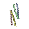
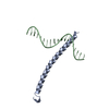
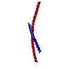
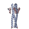
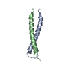
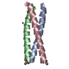
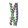
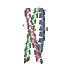
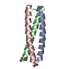
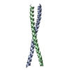
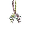
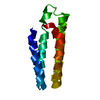
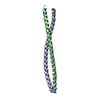
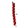
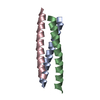
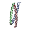
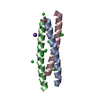
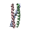
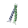
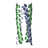
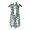
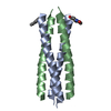
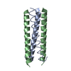
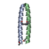
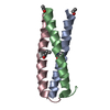
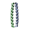
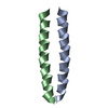
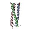
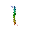
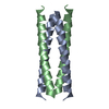
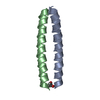
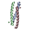
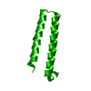
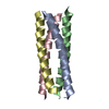
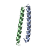
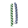
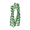
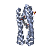
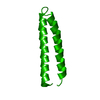
| Title | An anti-parallel four helix bundle (acetimide modification). | ||||||
 Components Components | GENERAL CONTROL PROTEIN GCN4 | ||||||
 Keywords Keywords | STRUCTURAL PROTEIN / FOUR HELIX BUNDLE / ANTIPARALLEL FOUR HELIX BUNDLE | ||||||
| Function / homology |  Function and homology information Function and homology informationFCERI mediated MAPK activation / protein localization to nuclear periphery / Activation of the AP-1 family of transcription factors / negative regulation of ribosomal protein gene transcription by RNA polymerase II / positive regulation of cellular response to amino acid starvation / response to amino acid starvation / mediator complex binding / Oxidative Stress Induced Senescence / amino acid biosynthetic process / TFIID-class transcription factor complex binding ...FCERI mediated MAPK activation / protein localization to nuclear periphery / Activation of the AP-1 family of transcription factors / negative regulation of ribosomal protein gene transcription by RNA polymerase II / positive regulation of cellular response to amino acid starvation / response to amino acid starvation / mediator complex binding / Oxidative Stress Induced Senescence / amino acid biosynthetic process / TFIID-class transcription factor complex binding / positive regulation of RNA polymerase II transcription preinitiation complex assembly / positive regulation of transcription initiation by RNA polymerase II / cellular response to nutrient levels / cellular response to amino acid starvation / RNA polymerase II transcription regulator complex / DNA-binding transcription activator activity, RNA polymerase II-specific / transcription regulator complex / sequence-specific DNA binding / RNA polymerase II-specific DNA-binding transcription factor binding / DNA-binding transcription factor activity, RNA polymerase II-specific / intracellular signal transduction / RNA polymerase II cis-regulatory region sequence-specific DNA binding / DNA-binding transcription factor activity / chromatin binding / negative regulation of transcription by RNA polymerase II / positive regulation of transcription by RNA polymerase II / identical protein binding / nucleus Similarity search - Function | ||||||
| Biological species |  | ||||||
| Method |  X-RAY DIFFRACTION / MOLECULAR REPLACEMENT / Resolution: 2.16 Å X-RAY DIFFRACTION / MOLECULAR REPLACEMENT / Resolution: 2.16 Å | ||||||
 Authors Authors | Yadav, M.K. / Leman, L.J. / Stout, C.D. / Ghadiri, M.R. | ||||||
 Citation Citation | Journal: Biochemistry / Year: 2005 Title: Structure-Based Engineering of Internal Cavities in Coiled-Coil Peptides Authors: Yadav, M.K. / Redman, J.E. / Leman, L.J. / Alvarez-Gutierrez, J.M. / Zhang, Y. / Stout, C.D. / Ghadiri, M.R. | ||||||
| History |
|
- Structure visualization
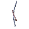
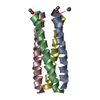
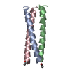
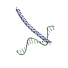
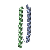
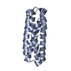
| Structure viewer | Molecule: MolmilJmol/JSmol |
|---|
- Downloads & links
-Download
| PDBx/mmCIF format | 1w5g.cif.gz | 24 KB | Display | PDBx/mmCIF format |
|---|---|---|---|---|
| PDB format | pdb1w5g.ent.gz | 16.2 KB | Display | PDB format |
| PDBx/mmJSON format | 1w5g.json.gz | Tree view | PDBx/mmJSON format | |
| Others |  Other downloads Other downloads |
-Validation report
| Arichive directory | https://data.pdbj.org/pub/pdb/validation_reports/w5/1w5gftp://data.pdbj.org/pub/pdb/validation_reports/w5/1w5g | HTTPS FTP |
|---|
-Related structure data
| Related structure data | 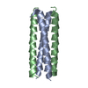 1untC 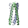 1unuC 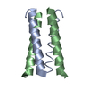 1unvC 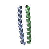 1unwC 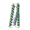 1unxC 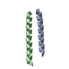 1unyC 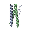 1unzC 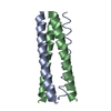 1uo0C 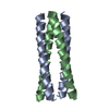 1uo1C 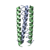 1uo2C 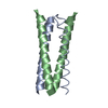 1uo3C  1uo4C 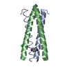 1uo5C 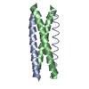 1w5iC 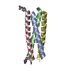 2bniC C: citing same article ( |
|---|---|
| Similar structure data |


-Links
 PDBj
PDBj

- Assembly
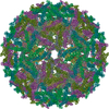
| Deposited unit | 
| ||||||||
|---|---|---|---|---|---|---|---|---|---|
| 1 | 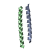
| ||||||||
| Unit cell |
|
-Components
| #1: Protein/peptide | Mass: 4020.829 Da / Num. of mol.: 2 / Mutation: YES / Source method: obtained synthetically Details: PEPTIDE REACTED WITH IODOACETIMIDE TO YEILD THIOETHER FROM CYSTEINE. NOTE SHIFT IN TYROSINE POSITION FROM PDB ENTRY 1VZL Source: (synth.) #2: Water | ChemComp-HOH / |  Mass: 18.015 Da / Num. of mol.: 2 / Source method: isolated from a natural source / Formula: H2O Mass: 18.015 Da / Num. of mol.: 2 / Source method: isolated from a natural source / Formula: H2OCompound details | CHAIN A, B ENGINEERED | |
|---|
-Experimental details
-Experiment
| Experiment | Method: X-RAY DIFFRACTION / Number of used crystals: 1 |
|---|
- Sample preparation
| Crystal | Density Matthews: 2 Å3/Da / Density % sol: 38.9 % |
|---|---|
| Crystal grow | Method: vapor diffusion, hanging drop / pH: 10 Details: HANGING DROP, 1UL OF 1MG/ML PEPTIDE IN WATER, 1UL 100MM CAPS, 30% PEG 400, PH 10.5. |
-Data collection
| Diffraction | Mean temperature: 93 K |
|---|---|
| Diffraction source | Source: ROTATING ANODE / Type: RIGAKU RU200 / Wavelength: 1.5418 |
| Detector | Date: May 29, 2004 / Details: CONFOCAL |
| Radiation | Monochromator: NI FILTER / Protocol: SINGLE WAVELENGTH / Monochromatic (M) / Laue (L): M / Scattering type: x-ray |
| Radiation wavelength | Wavelength: 1.5418 Å / Relative weight: 1 |
| Reflection | Resolution: 2.16→34.88 Å / Num. obs: 3960 / % possible obs: 100 % / Observed criterion σ(I): 2 / Redundancy: 5.37 % / Rmerge(I) obs: 0.05 / Net I/σ(I): 17.3 |
| Reflection shell | Resolution: 2.16→2.24 Å / Redundancy: 5.01 % / Rmerge(I) obs: 0.22 / Mean I/σ(I) obs: 6.5 / % possible all: 97.6 |
- Processing
| Software |
| ||||||||||||||||||||||||||||||||||||||||||||||||||||||||||||||||||||||||||||||||||||||||||||||||||||||||||||||||||||||||||||||||||||||||||||||||||||||||||||||||||||||||||||||||||||||
|---|---|---|---|---|---|---|---|---|---|---|---|---|---|---|---|---|---|---|---|---|---|---|---|---|---|---|---|---|---|---|---|---|---|---|---|---|---|---|---|---|---|---|---|---|---|---|---|---|---|---|---|---|---|---|---|---|---|---|---|---|---|---|---|---|---|---|---|---|---|---|---|---|---|---|---|---|---|---|---|---|---|---|---|---|---|---|---|---|---|---|---|---|---|---|---|---|---|---|---|---|---|---|---|---|---|---|---|---|---|---|---|---|---|---|---|---|---|---|---|---|---|---|---|---|---|---|---|---|---|---|---|---|---|---|---|---|---|---|---|---|---|---|---|---|---|---|---|---|---|---|---|---|---|---|---|---|---|---|---|---|---|---|---|---|---|---|---|---|---|---|---|---|---|---|---|---|---|---|---|---|---|---|---|
| Refinement | Method to determine structure: MOLECULAR REPLACEMENT / Resolution: 2.16→51.99 Å / Cor.coef. Fo:Fc: 0.881 / Cor.coef. Fo:Fc free: 0.853 / SU B: 6.182 / SU ML: 0.166 / Cross valid method: THROUGHOUT / ESU R: 0.351 / ESU R Free: 0.269 / Stereochemistry target values: MAXIMUM LIKELIHOOD / Details: HYDROGENS HAVE BEEN ADDED IN THE RIDING POSITIONS.
| ||||||||||||||||||||||||||||||||||||||||||||||||||||||||||||||||||||||||||||||||||||||||||||||||||||||||||||||||||||||||||||||||||||||||||||||||||||||||||||||||||||||||||||||||||||||
| Solvent computation | Ion probe radii: 0.8 Å / Shrinkage radii: 0.8 Å / VDW probe radii: 1.4 Å / Solvent model: BABINET MODEL WITH MASK | ||||||||||||||||||||||||||||||||||||||||||||||||||||||||||||||||||||||||||||||||||||||||||||||||||||||||||||||||||||||||||||||||||||||||||||||||||||||||||||||||||||||||||||||||||||||
| Displacement parameters | Biso mean: 40.43 Å2
| ||||||||||||||||||||||||||||||||||||||||||||||||||||||||||||||||||||||||||||||||||||||||||||||||||||||||||||||||||||||||||||||||||||||||||||||||||||||||||||||||||||||||||||||||||||||
| Refinement step | Cycle: LAST / Resolution: 2.16→51.99 Å
| ||||||||||||||||||||||||||||||||||||||||||||||||||||||||||||||||||||||||||||||||||||||||||||||||||||||||||||||||||||||||||||||||||||||||||||||||||||||||||||||||||||||||||||||||||||||
| Refine LS restraints |
|