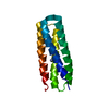
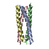
[English] 日本語
Yorodumi
- PDB-6ahx: Copper-Sensing Operon Regulator Protein (CsoRGz) -

+
Open data


ID or keywords:

Loading...

-
Basic information

Entry
Database: PDB / ID: 6ahx
TitleCopper-Sensing Operon Regulator Protein (CsoRGz)
ComponentsPutative cytosolic protein
KeywordsMETAL BINDING PROTEIN / Copper-Sensing Operon Regulator Protein
Function / homologyMetal-sensitive transcriptional repressor / Metal-sensitive repressor, helix protomer superfamily / Metal-sensitive transcriptional repressor / regulation of DNA-templated transcription / DNA binding / metal ion binding / Putative cytosolic protein
Function and homology information
Biological speciesGeobacillus zalihae (bacteria)
MethodX-RAY DIFFRACTION / MOLECULAR REPLACEMENT / Resolution: 2 Å
AuthorsNormi, M.Y. / Mangavelu, A. / Sayangku, A.A. / Jonet, M.A. / Adam, T.C.L. / Ali, M.S.M. / Rahman, R.N.Z.R.A. / Salleh, A.B.
Funding support Malaysia, 1items
OrganizationGrant numberCountry
Universiti Putra Malaysia Putra Postgraduate Initiative Grant9502200 Malaysia
CitationJournal: To Be Published
Title: Crystallization, Structural Determination and Analysis of Copper-sensing Operon Regulator Protein (CsoRGz) of Geobacillus zalihae Strain T1
Authors: Normi, M.Y. / Mangavelu, A. / Sayangku, A.A. / Jonet, M.A. / Adam, T.C.L. / Ali, M.S.M. / Rahman, R.N.Z.R.A. / Salleh, A.B.
History
DepositionAug 21, 2018Deposition site: PDBJ / Processing site: PDBJ
Revision 1.0Oct 23, 2019Provider: repository / Type: Initial release
Revision 1.1May 20, 2020Group: Author supporting evidence / Category: pdbx_audit_support / Item: _pdbx_audit_support.funding_organization
Revision 1.2Jul 1, 2020Group: Source and taxonomy / Category: entity_src_gen
Item: _entity_src_gen.pdbx_gene_src_gene / _entity_src_gen.pdbx_gene_src_ncbi_taxonomy_id / _entity_src_gen.pdbx_gene_src_scientific_name
Revision 1.3Nov 22, 2023Group: Data collection / Database references / Refinement description
Category: chem_comp_atom / chem_comp_bond ...chem_comp_atom / chem_comp_bond / database_2 / pdbx_initial_refinement_model
Item: _database_2.pdbx_DOI / _database_2.pdbx_database_accession

-
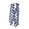
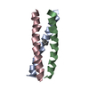
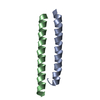
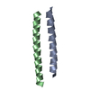
Structure visualization

Structure viewerMolecule:
MolmilJmol/JSmol

Downloads & links

-
Assembly

Deposited unit
A: Putative cytosolic protein


Theoretical massNumber of molelcules
Total (without water)11,7811
Polymers11,7811
Non-polymers00
Water86548
1
A: Putative cytosolic protein

A: Putative cytosolic protein


Theoretical massNumber of molelcules
Total (without water)23,5632
Polymers23,5632
Non-polymers00
Water362
TypeNameSymmetry operationNumber
identity operation1_555x,y,z1
crystal symmetry operation4_465y-1,x+1,-z1
Buried area2750 Å2
ΔGint-28 kcal/mol
Surface area6720 Å2
MethodPISA
Unit cell
Length a, b, c (Å)44.730, 44.730, 82.370
Angle α, β, γ (deg.)90.00, 90.00, 120.00
Int Tables number152
Space group name H-MP3121

-
Components

#1: Protein Putative cytosolic protein


Mass: 11781.420 Da / Num. of mol.: 1
Source method: isolated from a genetically manipulated source
Source: (gene. exp.) Geobacillus zalihae (bacteria) / Production host: Escherichia coli (E. coli) / References: UniProt: S7SWX2
#2: Water ChemComp-HOH / water


Mass: 18.015 Da / Num. of mol.: 48 / Source method: isolated from a natural source / Formula: H2O

-
Experimental details

-
Experiment

ExperimentMethod: X-RAY DIFFRACTION / Number of used crystals: 1

-
Sample preparation

CrystalDensity Matthews: 1.83 Å3/Da / Density % sol: 39.24 %
Crystal growTemperature: 293.95 K / Method: vapor diffusion, sitting drop / pH: 6.5
Details: 5% (v/v) Tacsimate TM pH 7.0, 0.1 M HEPES pH 7.0, 10% (w/v) PEG monomethyl ether 5000
PH range: 7

-
Data collection

DiffractionMean temperature: 100 K
Diffraction sourceSource: ROTATING ANODE / Type: Cu FINE FOCUS / Wavelength: 1.54 Å
DetectorType: DECTRIS PILATUS 200K / Detector: PIXEL / Date: Dec 6, 2016
RadiationProtocol: SINGLE WAVELENGTH / Monochromatic (M) / Laue (L): M / Scattering type: x-ray
Radiation wavelengthWavelength: 1.54 Å / Relative weight: 1
ReflectionResolution: 2→38.74 Å / Num. obs: 6882 / % possible obs: 100 % / Redundancy: 3.2 % / Rmerge(I) obs: 0.031 / Net I/σ(I): 45.7
Reflection shellResolution: 2→2.03 Å / Rmerge(I) obs: 0.028 / Num. unique obs: 664

-
Processing

Software
NameVersionClassification
REFMAC5.8.0103refinement
DENZOdata reduction
SCALEPACKdata scaling
MOLREPphasing
RefinementMethod to determine structure: MOLECULAR REPLACEMENT
Starting model: 3AAI
Resolution: 2→38.74 Å / Cor.coef. Fo:Fc: 0.956 / Cor.coef. Fo:Fc free: 0.924 / SU B: 5.568 / SU ML: 0.133 / Cross valid method: THROUGHOUT / ESU R: 0.145 / ESU R Free: 0.155 / Details: HYDROGENS HAVE BEEN ADDED IN THE RIDING POSITIONS
RfactorNum. reflection% reflectionSelection details
Rfree0.25796 345 5.3 %RANDOM
Rwork0.1967 ---
obs0.19996 6184 95.19 %-
Solvent computationIon probe radii: 0.8 Å / Shrinkage radii: 0.8 Å / VDW probe radii: 1.2 Å
Displacement parametersBiso mean: 35.033 Å2
Baniso -1Baniso -2Baniso -3
1-1.06 Å20.53 Å20 Å2
2--1.06 Å2-0 Å2
3----3.45 Å2
Refinement stepCycle: 1 / Resolution: 2→38.74 Å
ProteinNucleic acidLigandSolventTotal
Num. atoms471 0 0 48 519
Refine LS restraints
Refine-IDTypeDev idealDev ideal targetNumber
X-RAY DIFFRACTIONr_bond_refined_d0.0180.019475
X-RAY DIFFRACTIONr_bond_other_d0.0030.02495
X-RAY DIFFRACTIONr_angle_refined_deg1.9611.971635
X-RAY DIFFRACTIONr_angle_other_deg1.03631132
X-RAY DIFFRACTIONr_dihedral_angle_1_deg5.246559
X-RAY DIFFRACTIONr_dihedral_angle_2_deg28.00722.38121
X-RAY DIFFRACTIONr_dihedral_angle_3_deg18.6571596
X-RAY DIFFRACTIONr_dihedral_angle_4_deg22.046156
X-RAY DIFFRACTIONr_chiral_restr0.1030.274
X-RAY DIFFRACTIONr_gen_planes_refined0.0090.02521
X-RAY DIFFRACTIONr_gen_planes_other0.0010.02105
X-RAY DIFFRACTIONr_nbd_refined
X-RAY DIFFRACTIONr_nbd_other
X-RAY DIFFRACTIONr_nbtor_refined
X-RAY DIFFRACTIONr_nbtor_other
X-RAY DIFFRACTIONr_xyhbond_nbd_refined
X-RAY DIFFRACTIONr_xyhbond_nbd_other
X-RAY DIFFRACTIONr_metal_ion_refined
X-RAY DIFFRACTIONr_metal_ion_other
X-RAY DIFFRACTIONr_symmetry_vdw_refined
X-RAY DIFFRACTIONr_symmetry_vdw_other
X-RAY DIFFRACTIONr_symmetry_hbond_refined
X-RAY DIFFRACTIONr_symmetry_hbond_other
X-RAY DIFFRACTIONr_symmetry_metal_ion_refined
X-RAY DIFFRACTIONr_symmetry_metal_ion_other
X-RAY DIFFRACTIONr_mcbond_it2.5892.958239
X-RAY DIFFRACTIONr_mcbond_other2.5732.958238
X-RAY DIFFRACTIONr_mcangle_it3.6024.431297
X-RAY DIFFRACTIONr_mcangle_other3.64.432298
X-RAY DIFFRACTIONr_scbond_it5.2163.747236
X-RAY DIFFRACTIONr_scbond_other5.2053.747237
X-RAY DIFFRACTIONr_scangle_it
X-RAY DIFFRACTIONr_scangle_other8.3095.321339
X-RAY DIFFRACTIONr_long_range_B_refined9.79724.666580
X-RAY DIFFRACTIONr_long_range_B_other9.63824.222559
X-RAY DIFFRACTIONr_rigid_bond_restr
X-RAY DIFFRACTIONr_sphericity_free
X-RAY DIFFRACTIONr_sphericity_bonded
LS refinement shellResolution: 2→2.052 Å / Total num. of bins used: 20
RfactorNum. reflection% reflection
Rfree0.2 37 -
Rwork0.28 467 -
obs--98.05 %

+
About Yorodumi

-
News

-
Feb 9, 2022. New format data for meta-information of EMDB entries

New format data for meta-information of EMDB entries

  • Version 3 of the EMDB header file is now the official format.
  • The previous official version 1.9 will be removed from the archive.

Related info.:EMDB header

External links:wwPDB to switch to version 3 of the EMDB data model

-
Aug 12, 2020. Covid-19 info

Covid-19 info

URL: https://pdbjlvh1.pdbj.org/emnavi/covid19.php

New page: Covid-19 featured information page in EM Navigator.

Related info.:Covid-19 info / Mar 5, 2020. Novel coronavirus structure data

+
Mar 5, 2020. Novel coronavirus structure data

Novel coronavirus structure data

Related info.:Yorodumi Speices / Aug 12, 2020. Covid-19 info

External links:COVID-19 featured content - PDBj / Molecule of the Month (242):Coronavirus Proteases

+
Jan 31, 2019. EMDB accession codes are about to change! (news from PDBe EMDB page)

EMDB accession codes are about to change! (news from PDBe EMDB page)

  • The allocation of 4 digits for EMDB accession codes will soon come to an end. Whilst these codes will remain in use, new EMDB accession codes will include an additional digit and will expand incrementally as the available range of codes is exhausted. The current 4-digit format prefixed with “EMD-” (i.e. EMD-XXXX) will advance to a 5-digit format (i.e. EMD-XXXXX), and so on. It is currently estimated that the 4-digit codes will be depleted around Spring 2019, at which point the 5-digit format will come into force.
  • The EM Navigator/Yorodumi systems omit the EMD- prefix.

Related info.:Q: What is EMD? / ID/Accession-code notation in Yorodumi/EM Navigator

External links:EMDB Accession Codes are Changing Soon! / Contact to PDBj

+
Jul 12, 2017. Major update of PDB

Major update of PDB

  • wwPDB released updated PDB data conforming to the new PDBx/mmCIF dictionary.
  • This is a major update changing the version number from 4 to 5, and with Remediation, in which all the entries are updated.
  • In this update, many items about electron microscopy experimental information are reorganized (e.g. em_software).
  • Now, EM Navigator and Yorodumi are based on the updated data.

External links:wwPDB Remediation / Enriched Model Files Conforming to OneDep Data Standards Now Available in the PDB FTP Archive

-
Yorodumi

Thousand views of thousand structures

  • Yorodumi is a browser for structure data from EMDB, PDB, SASBDB, etc.
  • This page is also the successor to EM Navigator detail page, and also detail information page/front-end page for Omokage search.
  • The word "yorodu" (or yorozu) is an old Japanese word meaning "ten thousand". "mi" (miru) is to see.

Related info.:EMDB / PDB / SASBDB / Comparison of 3 databanks / Yorodumi Search / Aug 31, 2016. New EM Navigator & Yorodumi / Yorodumi Papers / Jmol/JSmol / Function and homology information / Changes in new EM Navigator and Yorodumi

Read more