 Movie
Movie Controller

[English] 日本語
 Yorodumi
- PDB-6hk5: X-ray structure of a truncated mutant of the metallochaperone Coo... -
+ Open data

- Basic information
| Entry | Database: PDB / ID: 6hk5 | ||||||
|---|---|---|---|---|---|---|---|
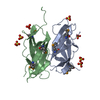
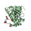
| Title | X-ray structure of a truncated mutant of the metallochaperone CooJ with a high-affinity nickel-binding site | ||||||
 Components Components | CooJ | ||||||
 Keywords Keywords | METAL BINDING PROTEIN / metallochaperone / nickel-binding | ||||||
| Function / homology | metal ion binding / NICKEL (II) ION / DI(HYDROXYETHYL)ETHER / TRIETHYLENE GLYCOL / 3,3',3''-phosphoryltripropanoic acid / CooJ Function and homology information Function and homology information | ||||||
| Biological species |  Rhodospirillum rubrum (bacteria) Rhodospirillum rubrum (bacteria) | ||||||
| Method |  X-RAY DIFFRACTION / SYNCHROTRON / SAD / Resolution: 2.042 Å X-RAY DIFFRACTION / SYNCHROTRON / SAD / Resolution: 2.042 Å | ||||||
 Authors Authors | Alfano, M. / Perard, J. / Basset, C. / Carpentier, P. / Zambelli, B. / Timm, J. / Crouzy, S. / Ciurli, S. / Cavazza, C. | ||||||
 Citation Citation | Journal: J.Biol.Chem. / Year: 2019 Title: The carbon monoxide dehydrogenase accessory protein CooJ is a histidine-rich multidomain dimer containing an unexpected Ni(II)-binding site. Authors: Alfano, M. / Perard, J. / Carpentier, P. / Basset, C. / Zambelli, B. / Timm, J. / Crouzy, S. / Ciurli, S. / Cavazza, C. | ||||||
| History |
|
- Structure visualization
| Structure viewer | Molecule: MolmilJmol/JSmol |
|---|
- Downloads & links
-Download
| PDBx/mmCIF format | 6hk5.cif.gz | 118.3 KB | Display | PDBx/mmCIF format |
|---|---|---|---|---|
| PDB format | pdb6hk5.ent.gz | 91 KB | Display | PDB format |
| PDBx/mmJSON format | 6hk5.json.gz | Tree view | PDBx/mmJSON format | |
| Others |  Other downloads Other downloads |
-Validation report
| Arichive directory | https://data.pdbj.org/pub/pdb/validation_reports/hk/6hk5ftp://data.pdbj.org/pub/pdb/validation_reports/hk/6hk5 | HTTPS FTP |
|---|
-Related structure data
| Similar structure data |
|---|


-Links
 PDBj
PDBj- Assembly
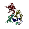
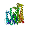
| Deposited unit | 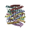
| ||||||||
|---|---|---|---|---|---|---|---|---|---|
| 1 | 
| ||||||||
| 2 | 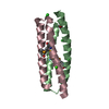
| ||||||||
| 3 | 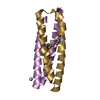
| ||||||||
| 4 | 
| ||||||||
| 5 | 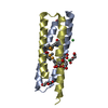
| ||||||||
| 6 | 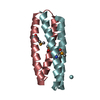
| ||||||||
| Unit cell |
|
-Components
-Protein , 1 types, 8 molecules BCEGADFH
| #1: Protein | Mass: 7606.244 Da / Num. of mol.: 8 / Fragment: Residues 1-68 Source method: isolated from a genetically manipulated source Source: (gene. exp.) Rhodospirillum rubrum (bacteria) / Gene: cooJ / Plasmid: pET15bProduction host: References: UniProt: P72321 |
|---|
-Non-polymers , 7 types, 314 molecules 


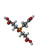

| #2: Chemical | ChemComp-NI /  Mass: 58.693 Da / Num. of mol.: 4 / Source method: obtained synthetically / Formula: Ni Mass: 58.693 Da / Num. of mol.: 4 / Source method: obtained synthetically / Formula: Ni#3: Chemical | ChemComp-PEG /  Mass: 106.120 Da / Num. of mol.: 4 / Source method: obtained synthetically / Formula: C4H10O3 Mass: 106.120 Da / Num. of mol.: 4 / Source method: obtained synthetically / Formula: C4H10O3#4: Chemical |  Mass: 35.453 Da / Num. of mol.: 3 / Source method: obtained synthetically / Formula: Cl Mass: 35.453 Da / Num. of mol.: 3 / Source method: obtained synthetically / Formula: Cl#5: Chemical | ChemComp-CA / |  Mass: 40.078 Da / Num. of mol.: 1 / Source method: obtained synthetically / Formula: Ca Mass: 40.078 Da / Num. of mol.: 1 / Source method: obtained synthetically / Formula: Ca#6: Chemical | ChemComp-PGE / |  Mass: 150.173 Da / Num. of mol.: 1 / Source method: obtained synthetically / Formula: C6H14O4 Mass: 150.173 Da / Num. of mol.: 1 / Source method: obtained synthetically / Formula: C6H14O4#7: Chemical | ChemComp-Z3P / |  Mass: 266.185 Da / Num. of mol.: 1 / Source method: obtained synthetically / Formula: C9H15O7P Mass: 266.185 Da / Num. of mol.: 1 / Source method: obtained synthetically / Formula: C9H15O7P#8: Water | ChemComp-HOH / | Mass: 18.015 Da / Num. of mol.: 300 / Source method: isolated from a natural source / Formula: H2O |
|---|
-Details
| Has protein modification | Y |
|---|
-Experimental details
-Experiment
| Experiment | Method: X-RAY DIFFRACTION / Number of used crystals: 1 |
|---|
- Sample preparation
| Crystal | Density Matthews: 2.05 Å3/Da / Density % sol: 40.1 % |
|---|---|
| Crystal grow | Temperature: 294 K / Method: vapor diffusion, hanging drop / pH: 7 Details: protein concentration of 10 mgmL-1 and 1 equivalent of Ni(II) per dimer, crystals were grown in 16% PEG3350 and 0.2 M calcium chloride |
-Data collection
| Diffraction | Mean temperature: 100 K |
|---|---|
| Diffraction source | Source: SYNCHROTRON / Site: ESRF  / Beamline: ID30B / Wavelength: 0.97856 Å / Beamline: ID30B / Wavelength: 0.97856 Å |
| Detector | Type: DECTRIS PILATUS3 6M / Detector: PIXEL / Date: Nov 12, 2017 |
| Radiation | Protocol: SINGLE WAVELENGTH / Monochromatic (M) / Laue (L): M / Scattering type: x-ray |
| Radiation wavelength | Wavelength: 0.97856 Å / Relative weight: 1 |
| Reflection | Resolution: 2.04→46.746 Å / Num. obs: 30586 / % possible obs: 99.2 % / Observed criterion σ(I): 1.5 / Redundancy: 6.4 % / Biso Wilson estimate: 31.4 Å2 / CC1/2: 0.997 / Rrim(I) all: 0.14 / Net I/σ(I): 9.4 |
| Reflection shell | Resolution: 2.04→2.16 Å / Redundancy: 6.2 % / Mean I/σ(I) obs: 1.63 / Num. unique obs: 4749 / CC1/2: 0.691 / Rrim(I) all: 0.88 / % possible all: 96 |
- Processing
| Software |
| |||||||||||||||||||||||||||||||||||||||||||||||||||||||||||||||||||||||||||||
|---|---|---|---|---|---|---|---|---|---|---|---|---|---|---|---|---|---|---|---|---|---|---|---|---|---|---|---|---|---|---|---|---|---|---|---|---|---|---|---|---|---|---|---|---|---|---|---|---|---|---|---|---|---|---|---|---|---|---|---|---|---|---|---|---|---|---|---|---|---|---|---|---|---|---|---|---|---|---|
| Refinement | Method to determine structure: SAD / Resolution: 2.042→46.746 Å / SU ML: 0.33 / Cross valid method: FREE R-VALUE / σ(F): 1.35 / Phase error: 29.3
| |||||||||||||||||||||||||||||||||||||||||||||||||||||||||||||||||||||||||||||
| Solvent computation | Shrinkage radii: 0.9 Å / VDW probe radii: 1.11 Å | |||||||||||||||||||||||||||||||||||||||||||||||||||||||||||||||||||||||||||||
| Refinement step | Cycle: LAST / Resolution: 2.042→46.746 Å
| |||||||||||||||||||||||||||||||||||||||||||||||||||||||||||||||||||||||||||||
| Refine LS restraints |
| |||||||||||||||||||||||||||||||||||||||||||||||||||||||||||||||||||||||||||||
| LS refinement shell |
|
