 Movie
Movie Controller


+ Open data

- Basic information
| Entry | Database: PDB / ID: 1p3q | ||||||
|---|---|---|---|---|---|---|---|
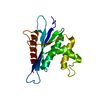
| Title | Mechanism of Ubiquitin Recognition by the CUE Domain of VPS9 | ||||||
 Components Components |
| ||||||
 Keywords Keywords | TRANSLATION / Trafficking / Post Translational Modification / Mono-Ubiquitination | ||||||
| Function / homology |  Function and homology information Function and homology information: / RAB GEFs exchange GTP for GDP on RABs / Translesion synthesis by REV1 / Recognition of DNA damage by PCNA-containing replication complex / Translesion Synthesis by POLH / Downregulation of ERBB4 signaling / Spry regulation of FGF signaling / Downregulation of ERBB2:ERBB3 signaling / NOD1/2 Signaling Pathway / APC/C:Cdc20 mediated degradation of Cyclin B ...: / RAB GEFs exchange GTP for GDP on RABs / Translesion synthesis by REV1 / Recognition of DNA damage by PCNA-containing replication complex / Translesion Synthesis by POLH / Downregulation of ERBB4 signaling / Spry regulation of FGF signaling / Downregulation of ERBB2:ERBB3 signaling / NOD1/2 Signaling Pathway / APC/C:Cdc20 mediated degradation of Cyclin B / APC-Cdc20 mediated degradation of Nek2A / EGFR downregulation / TCF dependent signaling in response to WNT / NRIF signals cell death from the nucleus / p75NTR recruits signalling complexes / NF-kB is activated and signals survival / Activated NOTCH1 Transmits Signal to the Nucleus / Downregulation of TGF-beta receptor signaling / TGF-beta receptor signaling in EMT (epithelial to mesenchymal transition) / Downregulation of SMAD2/3:SMAD4 transcriptional activity / SMAD2/SMAD3:SMAD4 heterotrimer regulates transcription / Senescence-Associated Secretory Phenotype (SASP) / activated TAK1 mediates p38 MAPK activation / JNK (c-Jun kinases) phosphorylation and activation mediated by activated human TAK1 / Regulation of FZD by ubiquitination / N-glycan trimming in the ER and Calnexin/Calreticulin cycle / Regulation of TNFR1 signaling / TNFR1-induced NF-kappa-B signaling pathway / Translesion synthesis by POLK / Translesion synthesis by POLI / Regulation of necroptotic cell death / HDR through Homologous Recombination (HRR) / Josephin domain DUBs / Recruitment and ATM-mediated phosphorylation of repair and signaling proteins at DNA double strand breaks / Processing of DNA double-strand break ends / Gap-filling DNA repair synthesis and ligation in GG-NER / Dual Incision in GG-NER / Fanconi Anemia Pathway / Regulation of TP53 Activity through Phosphorylation / Regulation of TP53 Degradation / Regulation of TP53 Activity through Methylation / Negative regulation of MET activity / PTK6 Regulates RTKs and Their Effectors AKT1 and DOK1 / Downregulation of ERBB2 signaling / E3 ubiquitin ligases ubiquitinate target proteins / Regulation of PTEN localization / ER Quality Control Compartment (ERQC) / Regulation of expression of SLITs and ROBOs / Interferon alpha/beta signaling / Endosomal Sorting Complex Required For Transport (ESCRT) / Activation of IRF3, IRF7 mediated by TBK1, IKKε (IKBKE) / IKK complex recruitment mediated by RIP1 / IRAK2 mediated activation of TAK1 complex / TRAF6-mediated induction of TAK1 complex within TLR4 complex / Alpha-protein kinase 1 signaling pathway / RAS processing / Pexophagy / Negative regulation of FLT3 / IRAK2 mediated activation of TAK1 complex upon TLR7/8 or 9 stimulation / Regulation of NF-kappa B signaling / Regulation of TBK1, IKKε (IKBKE)-mediated activation of IRF3, IRF7 / Regulation of pyruvate metabolism / SCF-beta-TrCP mediated degradation of Emi1 / MAP3K8 (TPL2)-dependent MAPK1/3 activation / Ovarian tumor domain proteases / Cyclin D associated events in G1 / Regulation of BACH1 activity / Regulation of innate immune responses to cytosolic DNA / Negative regulation of FGFR1 signaling / Negative regulation of FGFR2 signaling / Negative regulation of FGFR3 signaling / Negative regulation of FGFR4 signaling / Negative regulation of MAPK pathway / Formation of Incision Complex in GG-NER / Synthesis of active ubiquitin: roles of E1 and E2 enzymes / Inactivation of CSF3 (G-CSF) signaling / Termination of translesion DNA synthesis / Iron uptake and transport / Negative regulators of DDX58/IFIH1 signaling / Deactivation of the beta-catenin transactivating complex / Metalloprotease DUBs / vacuole inheritance / Formation of TC-NER Pre-Incision Complex / Dual incision in TC-NER / Gap-filling DNA repair synthesis and ligation in TC-NER / Autodegradation of Cdh1 by Cdh1:APC/C / APC/C:Cdc20 mediated degradation of Securin / APC/C:Cdh1 mediated degradation of Cdc20 and other APC/C:Cdh1 targeted proteins in late mitosis/early G1 / Cdc20:Phospho-APC/C mediated degradation of Cyclin A / Autodegradation of the E3 ubiquitin ligase COP1 / Asymmetric localization of PCP proteins / Degradation of AXIN / Degradation of DVL / Hedgehog ligand biogenesis / Hedgehog 'on' state / TNFR2 non-canonical NF-kB pathway / DNA Damage Recognition in GG-NER / Assembly of the pre-replicative complex / CDK-mediated phosphorylation and removal of Cdc6 / G2/M Checkpoints Similarity search - Function | ||||||
| Biological species |   | ||||||
| Method |  X-RAY DIFFRACTION / SYNCHROTRON / MAD / Resolution: 1.7 Å X-RAY DIFFRACTION / SYNCHROTRON / MAD / Resolution: 1.7 Å | ||||||
 Authors Authors | Prag, G. / Misra, S. / Jones, E.A. / Ghirlando, R. / Davies, B.A. / Horazdovsky, B.F. / Hurley, J.H. | ||||||
 Citation Citation | Journal: Cell(Cambridge,Mass.) / Year: 2003 Title: Mechanism of Ubiquitin Recognition by the CUE Domain of Vps9p. Authors: Prag, G. / Misra, S. / Jones, E.A. / Ghirlando, R. / Davies, B.A. / Horazdovsky, B.F. / Hurley, J.H. #1: Journal: Embo J. / Year: 2003Title: A ubiquitin-binding motif required for intramolecular monoubiquitylation, the CUE domain Authors: Shih, S.C. / Prag, G. / Francis, S.A. / Sutanto, M.A. / Hurley, J.H. / Hicke, L. | ||||||
| History |
|
- Structure visualization
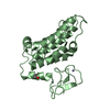
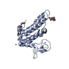
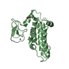
| Structure viewer | Molecule: MolmilJmol/JSmol |
|---|
- Downloads & links
-Download
| PDBx/mmCIF format | 1p3q.cif.gz | 60.9 KB | Display | PDBx/mmCIF format |
|---|---|---|---|---|
| PDB format | pdb1p3q.ent.gz | 44.8 KB | Display | PDB format |
| PDBx/mmJSON format | 1p3q.json.gz | Tree view | PDBx/mmJSON format | |
| Others |  Other downloads Other downloads |
-Validation report
| Arichive directory | https://data.pdbj.org/pub/pdb/validation_reports/p3/1p3qftp://data.pdbj.org/pub/pdb/validation_reports/p3/1p3q | HTTPS FTP |
|---|
-Related structure data


-Links
 PDBj
PDBj


- Assembly
| Deposited unit | 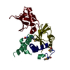
| ||||||||
|---|---|---|---|---|---|---|---|---|---|
| 1 | 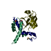
| ||||||||
| 2 | 
| ||||||||
| Unit cell |
|
-Components
| #1: Protein | Mass: 6034.562 Da / Num. of mol.: 2 / Fragment: CUE DOMAIN / Mutation: K435A, K436A Source method: isolated from a genetically manipulated source Source: (gene. exp.) Gene: VPS9 / Plasmid: pET-PARALLEL.HIS-CUE / Production host:  #2: Protein | Mass: 8576.831 Da / Num. of mol.: 2 / Source method: isolated from a natural source / Source: (natural) #3: Water | ChemComp-HOH / |  Mass: 18.015 Da / Num. of mol.: 176 / Source method: isolated from a natural source / Formula: H2O Mass: 18.015 Da / Num. of mol.: 176 / Source method: isolated from a natural source / Formula: H2OHas protein modification | Y | |
|---|
-Experimental details
-Experiment
| Experiment | Method: X-RAY DIFFRACTION / Number of used crystals: 1 |
|---|
- Sample preparation
| Crystal | Density Matthews: 2.44 Å3/Da / Density % sol: 49.23 % | |||||||||||||||||||||||||||||||||||||||||||||||||
|---|---|---|---|---|---|---|---|---|---|---|---|---|---|---|---|---|---|---|---|---|---|---|---|---|---|---|---|---|---|---|---|---|---|---|---|---|---|---|---|---|---|---|---|---|---|---|---|---|---|---|
| Crystal grow | Temperature: 293 K / Method: vapor diffusion, hanging drop / pH: 5.8 Details: PEG 3350, magnesium chloride, pH 5.8, VAPOR DIFFUSION, HANGING DROP, temperature 293K | |||||||||||||||||||||||||||||||||||||||||||||||||
| Crystal grow | *PLUS pH: 7.7 / Method: vapor diffusion, hanging drop | |||||||||||||||||||||||||||||||||||||||||||||||||
| Components of the solutions | *PLUS
|
-Data collection
| Diffraction | Mean temperature: 95 K | ||||||||||||
|---|---|---|---|---|---|---|---|---|---|---|---|---|---|
| Diffraction source | Source: SYNCHROTRON / Site: APS  / Beamline: 19-ID / Wavelength: 0.979174, 0.979311, 0.95645 / Beamline: 19-ID / Wavelength: 0.979174, 0.979311, 0.95645 | ||||||||||||
| Detector | Type: MARRESEARCH / Detector: CCD / Date: Oct 24, 2002 | ||||||||||||
| Radiation | Monochromator: Ni FILTER / Protocol: MAD / Monochromatic (M) / Laue (L): M / Scattering type: x-ray | ||||||||||||
| Radiation wavelength |
| ||||||||||||
| Reflection | Resolution: 1.7→50 Å / Num. all: 55378 / Num. obs: 28955 / % possible obs: 98.4 % / Observed criterion σ(F): 1 / Observed criterion σ(I): 2 / Redundancy: 3.5 % / Biso Wilson estimate: 22.1 Å2 / Rmerge(I) obs: 0.04 | ||||||||||||
| Reflection shell | Resolution: 1.7→1.76 Å / Rmerge(I) obs: 0.367 / Mean I/σ(I) obs: 2.3 / % possible all: 89.6 | ||||||||||||
| Reflection | *PLUS Highest resolution: 1.7 Å / Lowest resolution: 50 Å / Rmerge(I) obs: 0.04 | ||||||||||||
| Reflection shell | *PLUS Highest resolution: 1.7 Å / % possible obs: 89.6 % / Num. unique obs: 2626 |
- Processing
| Software |
| |||||||||||||||||||||||||
|---|---|---|---|---|---|---|---|---|---|---|---|---|---|---|---|---|---|---|---|---|---|---|---|---|---|---|
| Refinement | Method to determine structure: MAD / Resolution: 1.7→28.71 Å / Rfactor Rfree error: 0.005 / Isotropic thermal model: RESTRAINED / Cross valid method: THROUGHOUT / σ(F): 0 / Stereochemistry target values: Engh & Huber / Details: The structure was also refined with REFMAC 5.1
| |||||||||||||||||||||||||
| Solvent computation | Solvent model: FLAT MODEL / Bsol: 62.6478 Å2 / ksol: 0.366048 e/Å3 | |||||||||||||||||||||||||
| Displacement parameters | Biso mean: 29.1 Å2
| |||||||||||||||||||||||||
| Refine analyze |
| |||||||||||||||||||||||||
| Refinement step | Cycle: LAST / Resolution: 1.7→28.71 Å
| |||||||||||||||||||||||||
| Refine LS restraints |
| |||||||||||||||||||||||||
| LS refinement shell | Resolution: 1.7→1.81 Å / Rfactor Rfree error: 0.016 / Total num. of bins used: 6
| |||||||||||||||||||||||||
| Refinement | *PLUS Highest resolution: 1.7 Å / Lowest resolution: 50 Å / % reflection Rfree: 10 % / Rfactor Rwork: 0.26 | |||||||||||||||||||||||||
| Solvent computation | *PLUS | |||||||||||||||||||||||||
| Displacement parameters | *PLUS | |||||||||||||||||||||||||
| Refine LS restraints | *PLUS
|
