 Movie
Movie Controller

[English] 日本語
 Yorodumi
- PDB-3gr1: Periplasmic domain of the T3SS inner membrane protein PrgH from S... -
+ Open data

- Basic information
| Entry | Database: PDB / ID: 3gr1 | ||||||
|---|---|---|---|---|---|---|---|
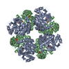
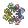
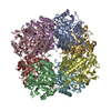
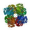
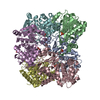
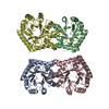
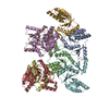
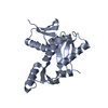
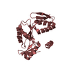
| Title | Periplasmic domain of the T3SS inner membrane protein PrgH from S.typhimurium (fragment 170-392) | ||||||
 Components Components | Protein prgH | ||||||
 Keywords Keywords | MEMBRANE PROTEIN / Type III secretion system / inner membrane protein / Cell membrane / Membrane / Transmembrane / Virulence | ||||||
| Function / homology |  Function and homology information Function and homology information | ||||||
| Biological species |  Salmonella typhimurium (bacteria) Salmonella typhimurium (bacteria) | ||||||
| Method |  X-RAY DIFFRACTION / SYNCHROTRON / MOLECULAR REPLACEMENT / Resolution: 2.8 Å X-RAY DIFFRACTION / SYNCHROTRON / MOLECULAR REPLACEMENT / Resolution: 2.8 Å | ||||||
 Authors Authors | Yip, C.K. / Vockovic, M. / Yu, A.C. / Strynadka, N.C.J. | ||||||
 Citation Citation | Journal: Nat.Struct.Mol.Biol. / Year: 2009 Title: A conserved structural motif mediates formation of the periplasmic rings in the type III secretion system. Authors: Spreter, T. / Yip, C.K. / Sanowar, S. / Andre, I. / Kimbrough, T.G. / Vuckovic, M. / Pfuetzner, R.A. / Deng, W. / Yu, A.C. / Finlay, B.B. / Baker, D. / Miller, S.I. / Strynadka, N.C. | ||||||
| History |
|
- Structure visualization
| Structure viewer | Molecule: MolmilJmol/JSmol |
|---|
- Downloads & links
-Download
| PDBx/mmCIF format | 3gr1.cif.gz | 310.5 KB | Display | PDBx/mmCIF format |
|---|---|---|---|---|
| PDB format | pdb3gr1.ent.gz | 255.4 KB | Display | PDB format |
| PDBx/mmJSON format | 3gr1.json.gz | Tree view | PDBx/mmJSON format | |
| Others |  Other downloads Other downloads |
-Validation report
| Arichive directory | https://data.pdbj.org/pub/pdb/validation_reports/gr/3gr1ftp://data.pdbj.org/pub/pdb/validation_reports/gr/3gr1 | HTTPS FTP |
|---|
-Related structure data
| Related structure data |  3gr0SC 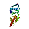 3gr5C S: Starting model for refinement C: citing same article ( |
|---|---|
| Similar structure data |
-Links
 PDBj
PDBj- Assembly


-Components
| #1: Protein | Mass: 26472.988 Da / Num. of mol.: 8 / Fragment: PrgH periplasmic domain (170-392) Source method: isolated from a genetically manipulated source Source: (gene. exp.) Salmonella typhimurium (bacteria) / Gene: prgH, STM2874 / Production host: |
|---|
-Experimental details
-Experiment
| Experiment | Method: X-RAY DIFFRACTION / Number of used crystals: 1 |
|---|
- Sample preparation
| Crystal | Density Matthews: 2.71 Å3/Da / Density % sol: 54.61 % |
|---|---|
| Crystal grow | Method: vapor diffusion, hanging drop / pH: 8 Details: 16% (w/v) PEG 3350 + 0.2 M tri-ammonium citrate + 0.1M Tris pH 8.0, VAPOR DIFFUSION, HANGING DROP |
-Data collection
| Diffraction source | Source: SYNCHROTRON / Site: ALS  / Beamline: 8.2.2 / Beamline: 8.2.2 |
|---|---|
| Detector | Type: ADSC QUANTUM 315 / Detector: CCD |
| Radiation | Protocol: SINGLE WAVELENGTH / Monochromatic (M) / Laue (L): M / Scattering type: x-ray |
| Radiation wavelength | Relative weight: 1 |
| Reflection | Resolution: 2.8→152.5 Å / Num. obs: 55188 / % possible obs: 96.5 % |
- Processing
| Software |
| ||||||||||||||||
|---|---|---|---|---|---|---|---|---|---|---|---|---|---|---|---|---|---|
| Refinement | Method to determine structure: MOLECULAR REPLACEMENT Starting model: 3GR0 Resolution: 2.8→152.5 Å / Cor.coef. Fo:Fc: 0.907 / Cor.coef. Fo:Fc free: 0.873 / SU ML: 0.326 / ESU R: 1.843 / ESU R Free: 0.417
| ||||||||||||||||
| Displacement parameters | Biso mean: 48.1 Å2
| ||||||||||||||||
| Refinement step | Cycle: LAST / Resolution: 2.8→152.5 Å
|
