 Movie
Movie Controller


+ Open data

- Basic information
| Entry | Database: PDB / ID: 1ixq | ||||||
|---|---|---|---|---|---|---|---|
| Title | Enzyme-Phosphate2 Complex of Pyridoxine 5'-Phosphate synthase | ||||||
 Components Components | Pyridoxine 5'-phosphate Synthase | ||||||
 Keywords Keywords | BIOSYNTHETIC PROTEIN / TIM barrel / enzyme-ligand complex / open-closed transition | ||||||
| Function / homology |  Function and homology information Function and homology informationpyridoxine 5'-phosphate synthase / pyridoxine 5'-phosphate synthase activity / pyridoxine biosynthetic process / identical protein binding / cytosol Similarity search - Function | ||||||
| Biological species |  | ||||||
| Method |  X-RAY DIFFRACTION / SIRAS / Resolution: 2.3 Å X-RAY DIFFRACTION / SIRAS / Resolution: 2.3 Å | ||||||
 Authors Authors | Garrido-Franco, M. / Laber, B. / Huber, R. / Clausen, T. | ||||||
 Citation Citation | Journal: J.MOL.BIOL. / Year: 2002 Title: Enzyme-ligand complexes of pyridoxine 5'-phosphate synthase: implications for substrate binding and catalysis Authors: Garrido-Franco, M. / Laber, B. / Huber, R. / Clausen, T. #1: Journal: Acta Crystallogr.,Sect.D / Year: 2000Title: Crystallization and preliminary X-ray crystallographic analysis of PdxJ, the pyridoxine 5'-phosphate synthesizing enzyme Authors: Garrido-Franco, M. / Huber, R. / Schmidt, F.S. / Laber, B. / Clausen, T. #2: Journal: Structure / Year: 2001Title: Structural Basis for the Function of Pyridoxine 5'-Phosphate Synthase Authors: Garrido-Franco, M. / Laber, B. / Huber, R. / Clausen, T. | ||||||
| History |
|
- Structure visualization
| Structure viewer | Molecule: MolmilJmol/JSmol |
|---|
- Downloads & links
-Download
| PDBx/mmCIF format | 1ixq.cif.gz | 189.7 KB | Display | PDBx/mmCIF format |
|---|---|---|---|---|
| PDB format | pdb1ixq.ent.gz | 152.8 KB | Display | PDB format |
| PDBx/mmJSON format | 1ixq.json.gz | Tree view | PDBx/mmJSON format | |
| Others |  Other downloads Other downloads |
-Validation report
| Arichive directory | https://data.pdbj.org/pub/pdb/validation_reports/ix/1ixqftp://data.pdbj.org/pub/pdb/validation_reports/ix/1ixq | HTTPS FTP |
|---|
-Related structure data


-Links
 PDBj
PDBj- Assembly
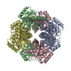
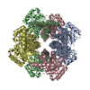
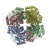
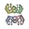
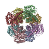
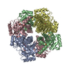
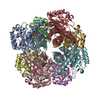
| Deposited unit | 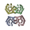
| ||||||||
|---|---|---|---|---|---|---|---|---|---|
| 1 | 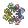
| ||||||||
| 2 | 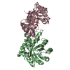
| ||||||||
| 3 | 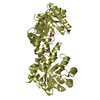
| ||||||||
| 4 | 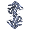
| ||||||||
| Unit cell |
| ||||||||
| Details | The biological assembly is an octamer generated from the tetramer in the asymmetric unit by the operations: x,-y,-z |
-Components
| #1: Protein | Mass: 26290.143 Da / Num. of mol.: 4 Source method: isolated from a genetically manipulated source Source: (gene. exp.) #2: Chemical | ChemComp-PO4 /   Mass: 94.971 Da / Num. of mol.: 4 / Source method: obtained synthetically / Formula: PO4 Mass: 94.971 Da / Num. of mol.: 4 / Source method: obtained synthetically / Formula: PO4#3: Water | ChemComp-HOH / |  Mass: 18.015 Da / Num. of mol.: 333 / Source method: isolated from a natural source / Formula: H2O Mass: 18.015 Da / Num. of mol.: 333 / Source method: isolated from a natural source / Formula: H2O |
|---|
-Experimental details
-Experiment
| Experiment | Method: X-RAY DIFFRACTION / Number of used crystals: 1 |
|---|
- Sample preparation
| Crystal | Density Matthews: 3.12 Å3/Da / Density % sol: 60.56 % | ||||||||||||||||||||||||||||||
|---|---|---|---|---|---|---|---|---|---|---|---|---|---|---|---|---|---|---|---|---|---|---|---|---|---|---|---|---|---|---|---|
| Crystal grow | Temperature: 293 K / Method: vapor diffusion, hanging drop / pH: 7.5 Details: 10% PEG6000, 2M NaCl, pH 7.5, VAPOR DIFFUSION, HANGING DROP, temperature 293K | ||||||||||||||||||||||||||||||
| Crystal grow | *PLUS pH: 8 / Method: vapor diffusion, sitting dropDetails: microseeding, Garrido-Franco, M., (2000) Acta Crystallogr., Sect.D, 56, 1045. | ||||||||||||||||||||||||||||||
| Components of the solutions | *PLUS
|
-Data collection
| Diffraction | Mean temperature: 100 K |
|---|---|
| Diffraction source | Source: ROTATING ANODE / Type: RIGAKU / Wavelength: 1.5418 Å |
| Detector | Type: MARRESEARCH / Detector: IMAGE PLATE / Date: Apr 5, 2001 |
| Radiation | Protocol: SINGLE WAVELENGTH / Monochromatic (M) / Laue (L): M / Scattering type: x-ray |
| Radiation wavelength | Wavelength: 1.5418 Å / Relative weight: 1 |
| Reflection | Resolution: 2.3→25 Å / Num. all: 198531 / Num. obs: 198531 / % possible obs: 99.8 % / Observed criterion σ(F): 0 / Observed criterion σ(I): 0 / Rmerge(I) obs: 0.045 / Rsym value: 0.045 / Net I/σ(I): 13.4 |
| Reflection shell | Resolution: 2.3→2.4 Å / Rmerge(I) obs: 0.343 / Mean I/σ(I) obs: 2.2 / % possible all: 99.2 |
| Reflection | *PLUS Num. obs: 58001 / Num. measured all: 198531 |
| Reflection shell | *PLUS % possible obs: 99.2 % |
- Processing
| Software |
| ||||||||||||||||||||
|---|---|---|---|---|---|---|---|---|---|---|---|---|---|---|---|---|---|---|---|---|---|
| Refinement | Method to determine structure: SIRAS Starting model: native enzyme Resolution: 2.3→20 Å / Isotropic thermal model: anisotropic / σ(F): 0 / σ(I): 0 / Stereochemistry target values: Engh & Huber
| ||||||||||||||||||||
| Refinement step | Cycle: LAST / Resolution: 2.3→20 Å
| ||||||||||||||||||||
| Refine LS restraints |
| ||||||||||||||||||||
| Refinement | *PLUS Lowest resolution: 25 Å / % reflection Rfree: 5 % / Rfactor Rfree: 0.27 / Rfactor Rwork: 0.221 | ||||||||||||||||||||
| Solvent computation | *PLUS | ||||||||||||||||||||
| Displacement parameters | *PLUS | ||||||||||||||||||||
| Refine LS restraints | *PLUS Type: c_angle_deg / Dev ideal: 1.46 |
