 Movie
Movie Controller

[English] 日本語
 Yorodumi
- PDB-1m5w: 1.96 A Crystal Structure of Pyridoxine 5'-Phosphate Synthase in C... -
+ Open data

- Basic information
| Entry | Database: PDB / ID: 1m5w | ||||||
|---|---|---|---|---|---|---|---|
| Title | 1.96 A Crystal Structure of Pyridoxine 5'-Phosphate Synthase in Complex with 1-deoxy-D-xylulose phosphate | ||||||
 Components Components | Pyridoxal phosphate biosynthetic protein pdxJ | ||||||
 Keywords Keywords | BIOSYNTHETIC PROTEIN / TIM barrel / protein-substrate complex / multi-binding states | ||||||
| Function / homology |  Function and homology information Function and homology informationpyridoxine 5'-phosphate synthase / pyridoxine 5'-phosphate synthase activity / pyridoxine biosynthetic process / identical protein binding / cytosol Similarity search - Function | ||||||
| Biological species |  | ||||||
| Method |  X-RAY DIFFRACTION / SYNCHROTRON / MOLECULAR REPLACEMENT / Resolution: 1.96 Å X-RAY DIFFRACTION / SYNCHROTRON / MOLECULAR REPLACEMENT / Resolution: 1.96 Å | ||||||
 Authors Authors | Yeh, J.I. / Du, S. / Pohl, E. / Cane, D.E. | ||||||
 Citation Citation | Journal: Biochemistry / Year: 2002 Title: Multistate Binding in Pyridoxine 5'-Phosphate Synthase: 1.96 A Crystal Structure in Complex with 1-deoxy-D-xylulose phosphate Authors: Yeh, J.I. / Du, S. / Pohl, E. / Cane, D.E. | ||||||
| History |
|
- Structure visualization
| Structure viewer | Molecule: MolmilJmol/JSmol |
|---|
- Downloads & links
-Download
| PDBx/mmCIF format | 1m5w.cif.gz | 380.4 KB | Display | PDBx/mmCIF format |
|---|---|---|---|---|
| PDB format | pdb1m5w.ent.gz | 312.9 KB | Display | PDB format |
| PDBx/mmJSON format | 1m5w.json.gz | Tree view | PDBx/mmJSON format | |
| Others |  Other downloads Other downloads |
-Validation report
| Arichive directory | https://data.pdbj.org/pub/pdb/validation_reports/m5/1m5wftp://data.pdbj.org/pub/pdb/validation_reports/m5/1m5w | HTTPS FTP |
|---|
-Related structure data
| Related structure data |  1ho4S S: Starting model for refinement |
|---|---|
| Similar structure data |

-Links
 PDBj
PDBj- Assembly
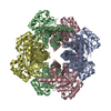
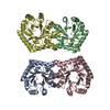
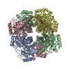
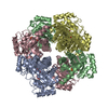
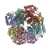
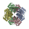
| Deposited unit | 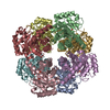
| ||||||||
|---|---|---|---|---|---|---|---|---|---|
| 1 |
| ||||||||
| 2 | 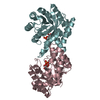
| ||||||||
| 3 | 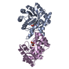
| ||||||||
| 4 | 
| ||||||||
| 5 | 
| ||||||||
| Unit cell |
|
-Components
| #1: Protein | Mass: 26421.340 Da / Num. of mol.: 8 Source method: isolated from a genetically manipulated source Source: (gene. exp.) #2: Chemical | ChemComp-DXP / 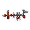  Mass: 214.110 Da / Num. of mol.: 6 Mass: 214.110 Da / Num. of mol.: 6Source method: isolated from a genetically manipulated source Formula: C5H11O7P #3: Chemical |   Mass: 94.971 Da / Num. of mol.: 2 / Source method: obtained synthetically / Formula: PO4 Mass: 94.971 Da / Num. of mol.: 2 / Source method: obtained synthetically / Formula: PO4#4: Water | ChemComp-HOH / |  Mass: 18.015 Da / Num. of mol.: 943 / Source method: isolated from a natural source / Formula: H2O Mass: 18.015 Da / Num. of mol.: 943 / Source method: isolated from a natural source / Formula: H2O |
|---|
-Experimental details
-Experiment
| Experiment | Method: X-RAY DIFFRACTION / Number of used crystals: 2 |
|---|
- Sample preparation
| Crystal | Density Matthews: 2.69 Å3/Da / Density % sol: 54.35 % | |||||||||||||||||||||||||
|---|---|---|---|---|---|---|---|---|---|---|---|---|---|---|---|---|---|---|---|---|---|---|---|---|---|---|
| Crystal grow | Temperature: 297 K / Method: vapor diffusion, hanging drop / pH: 7 Details: PEG 8000, PEG 1000, glycerol, pH 7, VAPOR DIFFUSION, HANGING DROP, temperature 297K | |||||||||||||||||||||||||
| Crystal grow | *PLUS Method: vapor diffusion, hanging drop | |||||||||||||||||||||||||
| Components of the solutions | *PLUS
|
-Data collection
| Diffraction | Mean temperature: 100 K |
|---|---|
| Diffraction source | Source: SYNCHROTRON / Site: EMBL/DESY, HAMBURG  / Beamline: X11 / Wavelength: 1.05 Å / Beamline: X11 / Wavelength: 1.05 Å |
| Detector | Type: MARRESEARCH / Detector: CCD / Date: Aug 21, 2000 |
| Radiation | Monochromator: wiggler / Protocol: SINGLE WAVELENGTH / Monochromatic (M) / Laue (L): M / Scattering type: x-ray |
| Radiation wavelength | Wavelength: 1.05 Å / Relative weight: 1 |
| Reflection | Resolution: 1.96→50 Å / Num. all: 163858 / Num. obs: 157249 / % possible obs: 96 % / Observed criterion σ(F): 3 / Observed criterion σ(I): 3 / Redundancy: 4.7 % / Biso Wilson estimate: 20.4 Å2 / Rmerge(I) obs: 0.045 / Rsym value: 0.045 / Net I/σ(I): 18.8 |
| Reflection shell | Resolution: 1.96→2.03 Å / Redundancy: 3.7 % / Rmerge(I) obs: 0.254 / Mean I/σ(I) obs: 2.6 / Num. unique all: 13719 / Rsym value: 0.304 / % possible all: 84.4 |
| Reflection | *PLUS Lowest resolution: 20 Å / % possible obs: 96 % / Num. measured all: 494561 |
| Reflection shell | *PLUS % possible obs: 84.4 % |
- Processing
| Software |
| |||||||||||||||||||||||||
|---|---|---|---|---|---|---|---|---|---|---|---|---|---|---|---|---|---|---|---|---|---|---|---|---|---|---|
| Refinement | Method to determine structure: MOLECULAR REPLACEMENT Starting model: PDB ENTRY 1HO4 Resolution: 1.96→6 Å / Isotropic thermal model: isotropic / Cross valid method: THROUGHOUT / σ(F): 0 / σ(I): 0 / Stereochemistry target values: Engh & Huber
| |||||||||||||||||||||||||
| Displacement parameters | Biso mean: 20.4 Å2 | |||||||||||||||||||||||||
| Refine analyze |
| |||||||||||||||||||||||||
| Refinement step | Cycle: LAST / Resolution: 1.96→6 Å
| |||||||||||||||||||||||||
| Refine LS restraints |
| |||||||||||||||||||||||||
| Refinement | *PLUS % reflection Rfree: 10 % | |||||||||||||||||||||||||
| Solvent computation | *PLUS | |||||||||||||||||||||||||
| Displacement parameters | *PLUS | |||||||||||||||||||||||||
| Refine LS restraints | *PLUS
|
