 Movie
Movie Controller

[English] 日本語
 Yorodumi
- PDB-1ho4: CRYSTAL STRUCTURE OF PYRIDOXINE 5'-PHOSPHATE SYNTHASE IN COMPLEX ... -
+ Open data

- Basic information
| Entry | Database: PDB / ID: 1ho4 | ||||||
|---|---|---|---|---|---|---|---|
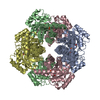
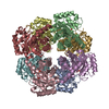
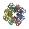
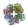
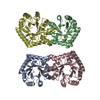
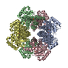
| Title | CRYSTAL STRUCTURE OF PYRIDOXINE 5'-PHOSPHATE SYNTHASE IN COMPLEX WITH PYRIDOXINE 5'-PHOSPHATE AND INORGANIC PHOSPHATE | ||||||
 Components Components | PYRIDOXINE 5'-PHOSPHATE SYNTHASE | ||||||
 Keywords Keywords | BIOSYNTHETIC PROTEIN / TIM Barrel / Open-Closed Transition / Enzyme-Product Complex / Water Channel | ||||||
| Function / homology |  Function and homology information Function and homology informationpyridoxine 5'-phosphate synthase / pyridoxine 5'-phosphate synthase activity / pyridoxine biosynthetic process / identical protein binding / cytosol Similarity search - Function | ||||||
| Biological species |  | ||||||
| Method |  X-RAY DIFFRACTION / MOLECULAR REPLACEMENT / Resolution: 2.3 Å X-RAY DIFFRACTION / MOLECULAR REPLACEMENT / Resolution: 2.3 Å | ||||||
 Authors Authors | Garrido-Franco, M. / Laber, B. / Huber, R. / Clausen, T. | ||||||
 Citation Citation | Journal: Structure / Year: 2001 Title: Structural basis for the function of pyridoxine 5'-phosphate synthase. Authors: Franco, M.G. / Laber, B. / Huber, R. / Clausen, T. | ||||||
| History |
|
- Structure visualization
| Structure viewer | Molecule: MolmilJmol/JSmol |
|---|
- Downloads & links
-Download
| PDBx/mmCIF format | 1ho4.cif.gz | 206.3 KB | Display | PDBx/mmCIF format |
|---|---|---|---|---|
| PDB format | pdb1ho4.ent.gz | 165.3 KB | Display | PDB format |
| PDBx/mmJSON format | 1ho4.json.gz | Tree view | PDBx/mmJSON format | |
| Others |  Other downloads Other downloads |
-Validation report
| Arichive directory | https://data.pdbj.org/pub/pdb/validation_reports/ho/1ho4ftp://data.pdbj.org/pub/pdb/validation_reports/ho/1ho4 | HTTPS FTP |
|---|
-Related structure data
| Related structure data |  1ho1C  1hoiS C: citing same article ( S: Starting model for refinement |
|---|---|
| Similar structure data |
-Links
 PDBj
PDBj- Assembly
| Deposited unit | 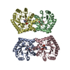
| ||||||||
|---|---|---|---|---|---|---|---|---|---|
| 1 | 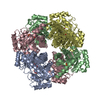
| ||||||||
| 2 | 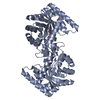
| ||||||||
| 3 | 
| ||||||||
| 4 | 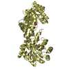
| ||||||||
| Unit cell |
| ||||||||
| Details | The biological assembly is an octamer generated from the tetramer in the asymmetric unit by the operations: x,-y,-z |
-Components
| #1: Protein | Mass: 26290.143 Da / Num. of mol.: 4 Source method: isolated from a genetically manipulated source Source: (gene. exp.) #2: Chemical |   Mass: 94.971 Da / Num. of mol.: 3 / Source method: obtained synthetically / Formula: PO4 Mass: 94.971 Da / Num. of mol.: 3 / Source method: obtained synthetically / Formula: PO4#3: Chemical | ChemComp-PXP /   Mass: 249.158 Da / Num. of mol.: 4 / Source method: obtained synthetically / Formula: C8H12NO6P Mass: 249.158 Da / Num. of mol.: 4 / Source method: obtained synthetically / Formula: C8H12NO6P#4: Water | ChemComp-HOH / |  Mass: 18.015 Da / Num. of mol.: 735 / Source method: isolated from a natural source / Formula: H2O Mass: 18.015 Da / Num. of mol.: 735 / Source method: isolated from a natural source / Formula: H2O |
|---|
-Experimental details
-Experiment
| Experiment | Method: X-RAY DIFFRACTION / Number of used crystals: 1 |
|---|
- Sample preparation
| Crystal | Density Matthews: 3.05 Å3/Da / Density % sol: 59.68 % |
|---|---|
| Crystal grow | Temperature: 293 K / Method: vapor diffusion, hanging drop / pH: 7.5 Details: 10% PEG6000, 2M NaCl, pH 7.5, VAPOR DIFFUSION, HANGING DROP, temperature 20K |
-Data collection
| Diffraction | Mean temperature: 100 K |
|---|---|
| Diffraction source | Source: ROTATING ANODE / Type: RIGAKU RU300 / Wavelength: 1.5418 Å |
| Detector | Type: MARRESEARCH / Detector: IMAGE PLATE / Date: Jan 31, 2000 |
| Radiation | Protocol: SINGLE WAVELENGTH / Monochromatic (M) / Laue (L): M / Scattering type: x-ray |
| Radiation wavelength | Wavelength: 1.5418 Å / Relative weight: 1 |
| Reflection | Resolution: 2→25 Å / Num. all: 54860 / Num. obs: 110743 / % possible obs: 96.2 % / Observed criterion σ(F): 0 / Observed criterion σ(I): 0 / Redundancy: 2 % / Biso Wilson estimate: 38.8 Å2 / Rmerge(I) obs: 0.046 / Rsym value: 0.046 / Net I/σ(I): 8.6 |
| Reflection shell | Resolution: 2.3→2.4 Å / Redundancy: 2.4 % / Rmerge(I) obs: 0.208 / Mean I/σ(I) obs: 3.6 / Num. unique all: 13071 / Rsym value: 0.208 / % possible all: 95.7 |
| Reflection | *PLUS Num. obs: 54860 / Num. measured all: 110743 |
| Reflection shell | *PLUS % possible obs: 95.7 % |
- Processing
| Software |
| ||||||||||||||||||||
|---|---|---|---|---|---|---|---|---|---|---|---|---|---|---|---|---|---|---|---|---|---|
| Refinement | Method to determine structure: MOLECULAR REPLACEMENT Starting model: PDB entry 1HOI Resolution: 2.3→20 Å / Isotropic thermal model: Anisotropic / σ(F): 0 / σ(I): 0 / Stereochemistry target values: Engh and Huber
| ||||||||||||||||||||
| Displacement parameters | Biso mean: 56.6 Å2 | ||||||||||||||||||||
| Refinement step | Cycle: LAST / Resolution: 2.3→20 Å
| ||||||||||||||||||||
| Refine LS restraints |
| ||||||||||||||||||||
| Software | *PLUS Name: CNS / Version: 1 / Classification: refinement | ||||||||||||||||||||
| Refine LS restraints | *PLUS Type: c_bond_d |
