[English] 日本語
Yorodumi
- PDB-1e51: Crystal structure of native human erythrocyte 5-aminolaevulinic a... -

+
Open data


ID or keywords:

Loading...

-
Basic information

Entry
Database: PDB / ID: 1.0E+51
Title
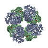
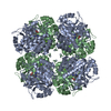
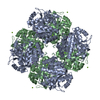
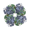
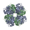
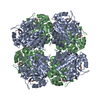
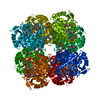
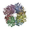
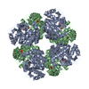
Crystal structure of native human erythrocyte 5-aminolaevulinic acid dehydratase
ComponentsDELTA-AMINOLEVULINIC ACID DEHYDRATASE
KeywordsLYASE / DEHYDRATASE / TETRAPYRROLE BIOSYNTHESIS / TIM BARREL / PORPHOBILINOGEN SYNTHASE / LEAD POISONING
Function / homology
Function and homology information


proteasome core complex binding / response to vitamin B1 / response to platinum ion / porphobilinogen synthase / porphobilinogen synthase activity / heme B biosynthetic process / heme O biosynthetic process / heme A biosynthetic process / negative regulation of proteasomal protein catabolic process / cellular response to lead ion ...proteasome core complex binding / response to vitamin B1 / response to platinum ion / porphobilinogen synthase / porphobilinogen synthase activity / heme B biosynthetic process / heme O biosynthetic process / heme A biosynthetic process / negative regulation of proteasomal protein catabolic process / cellular response to lead ion / response to mercury ion / response to aluminum ion / response to selenium ion / protoporphyrinogen IX biosynthetic process / response to fatty acid / response to cobalt ion / response to methylmercury / response to arsenic-containing substance / response to iron ion / response to herbicide / Heme biosynthesis / heme biosynthetic process / response to ionizing radiation / response to zinc ion / response to vitamin E / response to amino acid / response to cadmium ion / catalytic activity / response to glucocorticoid / cellular response to interleukin-4 / response to activity / protein homooligomerization / response to ethanol / secretory granule lumen / response to oxidative stress / ficolin-1-rich granule lumen / response to lipopolysaccharide / response to hypoxia / response to xenobiotic stimulus / Neutrophil degranulation / extracellular exosome / zinc ion binding / extracellular region / identical protein binding / nucleus / cytosol
Similarity search - Function
Delta-aminolevulinic acid dehydratase / Delta-aminolevulinic acid dehydratase, active site / Delta-aminolevulinic acid dehydratase / Delta-aminolevulinic acid dehydratase active site. / Delta-aminolevulinic acid dehydratase / Aldolase class I / Aldolase-type TIM barrel / TIM Barrel / Alpha-Beta Barrel / Alpha Beta
Similarity search - Domain/homology
Chem-PBG / Delta-aminolevulinic acid dehydratase
Similarity search - Component
Biological speciesHOMO SAPIENS (human)
MethodX-RAY DIFFRACTION / SYNCHROTRON / MOLECULAR REPLACEMENT / Resolution: 2.83 Å
AuthorsMills-Davies, N.L. / Thompson, D. / Cooper, J.B. / Shoolingin-Jordan, P.M.
CitationJournal: To be Published
Title: The Crystal Structure of Human Ala-Dehydratase
Authors: Mills-Davies, N.L. / Thompson, D. / Cooper, J.B. / Wood, S.P. / Shoolingin-Jordan, P.M.
History
DepositionJul 13, 2000Deposition site: PDBE / Processing site: PDBE
Revision 1.0Jul 12, 2001Provider: repository / Type: Initial release
Revision 1.1Feb 5, 2014Group: Data collection / Database references ...Data collection / Database references / Derived calculations / Other / Refinement description / Structure summary / Version format compliance
Revision 1.2Apr 18, 2018Group: Data collection / Database references / Category: citation
Revision 1.3Dec 13, 2023Group: Data collection / Database references ...Data collection / Database references / Derived calculations / Other / Refinement description
Category: chem_comp_atom / chem_comp_bond ...chem_comp_atom / chem_comp_bond / database_2 / pdbx_database_status / pdbx_initial_refinement_model / pdbx_struct_conn_angle / struct_conn / struct_site
Item: _database_2.pdbx_DOI / _database_2.pdbx_database_accession ..._database_2.pdbx_DOI / _database_2.pdbx_database_accession / _pdbx_database_status.status_code_sf / _pdbx_struct_conn_angle.ptnr1_auth_comp_id / _pdbx_struct_conn_angle.ptnr1_auth_seq_id / _pdbx_struct_conn_angle.ptnr1_label_asym_id / _pdbx_struct_conn_angle.ptnr1_label_atom_id / _pdbx_struct_conn_angle.ptnr1_label_comp_id / _pdbx_struct_conn_angle.ptnr1_label_seq_id / _pdbx_struct_conn_angle.ptnr3_auth_comp_id / _pdbx_struct_conn_angle.ptnr3_auth_seq_id / _pdbx_struct_conn_angle.ptnr3_label_asym_id / _pdbx_struct_conn_angle.ptnr3_label_atom_id / _pdbx_struct_conn_angle.ptnr3_label_comp_id / _pdbx_struct_conn_angle.ptnr3_label_seq_id / _pdbx_struct_conn_angle.value / _struct_conn.pdbx_dist_value / _struct_conn.pdbx_leaving_atom_flag / _struct_conn.ptnr1_auth_comp_id / _struct_conn.ptnr1_auth_seq_id / _struct_conn.ptnr1_label_asym_id / _struct_conn.ptnr1_label_atom_id / _struct_conn.ptnr1_label_comp_id / _struct_conn.ptnr1_label_seq_id / _struct_conn.ptnr2_auth_comp_id / _struct_conn.ptnr2_auth_seq_id / _struct_conn.ptnr2_label_asym_id / _struct_conn.ptnr2_label_atom_id / _struct_conn.ptnr2_label_comp_id / _struct_conn.ptnr2_label_seq_id / _struct_site.pdbx_auth_asym_id / _struct_site.pdbx_auth_comp_id / _struct_site.pdbx_auth_seq_id

-
Structure visualization

Structure viewerMolecule:
MolmilJmol/JSmol

Downloads & links

-
Assembly

Deposited unit
A: DELTA-AMINOLEVULINIC ACID DEHYDRATASE
B: DELTA-AMINOLEVULINIC ACID DEHYDRATASE
hetero molecules


Theoretical massNumber of molelcules
Total (without water)73,1616
Polymers72,6782
Non-polymers4844
Water0
1
A: DELTA-AMINOLEVULINIC ACID DEHYDRATASE
B: DELTA-AMINOLEVULINIC ACID DEHYDRATASE
hetero molecules

A: DELTA-AMINOLEVULINIC ACID DEHYDRATASE
B: DELTA-AMINOLEVULINIC ACID DEHYDRATASE
hetero molecules

A: DELTA-AMINOLEVULINIC ACID DEHYDRATASE
B: DELTA-AMINOLEVULINIC ACID DEHYDRATASE
hetero molecules

A: DELTA-AMINOLEVULINIC ACID DEHYDRATASE
B: DELTA-AMINOLEVULINIC ACID DEHYDRATASE
hetero molecules


Theoretical massNumber of molelcules
Total (without water)292,64624
Polymers290,7118
Non-polymers1,93516
Water0
TypeNameSymmetry operationNumber
identity operation1_555x,y,z1
crystal symmetry operation4_565y,-x+1,z1
crystal symmetry operation3_655-y+1,x,z1
crystal symmetry operation2_665-x+1,-y+1,z1
Buried area43900 Å2
ΔGint-509.2 kcal/mol
Surface area72950 Å2
MethodPISA
Unit cell
Length a, b, c (Å)125.530, 125.530, 200.910
Angle α, β, γ (deg.)90.00, 90.00, 90.00
Int Tables number97
Space group name H-MI422

-
Components

#1: Protein DELTA-AMINOLEVULINIC ACID DEHYDRATASE / / ALADH / PORPHOBILINOGEN SYNTHASE / PORPHOBILINOGEN SYNTHASE / ALADH


Mass: 36338.852 Da / Num. of mol.: 2 / Source method: isolated from a natural source / Details: PORPHOBILINOGEN IN ACTIVE SITE OF MONOMER A / Source: (natural) HOMO SAPIENS (human) / Cell: ERYTHROCYTES / References: UniProt: P13716, porphobilinogen synthase
#2: Chemical ChemComp-PBG / 3-[5-(AMINOMETHYL)-4-(CARBOXYMETHYL)-1H-PYRROL-3-YL]PROPANOIC ACID / 2-AMINOMETHYLPYRROL-3-ACETIC ACID 4-PROPIONIC ACID / PORPHOBILINOGEN / 5-(AMINOMETHYL)-4-(CARBOXYMETHYL)-1H-PYRROLE-3-PROPANOIC ACID / Porphobilinogen


Mass: 226.229 Da / Num. of mol.: 1 / Source method: obtained synthetically / Formula: C10H14N2O4
#3: Chemical ChemComp-ZN / ZINC ION


Mass: 65.409 Da / Num. of mol.: 1 / Source method: obtained synthetically / Formula: Zn
#4: Chemical ChemComp-SO4 / SULFATE ION / Sulfate


Mass: 96.063 Da / Num. of mol.: 2 / Source method: obtained synthetically / Formula: SO4
Compound detailsPORPHOBILINOGEN PRODUCT MOLECULE BOUND AT MONOMER A ACTIVE SITE. SECOND STEP IN PORPHYRIN AND HEME ...PORPHOBILINOGEN PRODUCT MOLECULE BOUND AT MONOMER A ACTIVE SITE. SECOND STEP IN PORPHYRIN AND HEME BIOSYNTHESIS DISEASE: DEFECTS IN ALAD ARE THE CAUSE OF ACUTE HEPATIC PORPHYRIA CATALYTIC ACTIVITY: 2 5-AMINOLEVULINATE = PORPHOBILINOGEN + H(2)O COFACTOR: ZINC POLYMORPHISM: THERE ARE TWO COMMON ALLELES OF ALAD. INDIVIDUALS HETEROZYGOUS OR HOMOZYGOUS FOR THE 2ND ALLELE HAVE SIGNIFICANTLY HIGHER BLOOD LEAD LEVELS THAN DO 1ST ALLELE HOMOZYGOTES WHEN EXPOSED TO ENVIRONMENTAL LEAD.

-
Experimental details

-
Experiment

ExperimentMethod: X-RAY DIFFRACTION / Number of used crystals: 1

-
Sample preparation

CrystalDensity Matthews: 2.72 Å3/Da / Density % sol: 56 %
Crystal growpH: 6.2 / Details: MES, AMMONIUM SULPHATE, DIOXANE, pH 6.20

-
Data collection

DiffractionMean temperature: 100 K
Diffraction sourceSource: SYNCHROTRON / Site: SRS / Beamline: PX9.5 / Wavelength: 0.87
DetectorType: MARRESEARCH / Detector: IMAGE PLATE / Date: Apr 15, 1998
RadiationProtocol: SINGLE WAVELENGTH / Monochromatic (M) / Laue (L): M / Scattering type: x-ray
Radiation wavelengthWavelength: 0.87 Å / Relative weight: 1
ReflectionResolution: 2.832→54.233 Å / Num. obs: 18652 / % possible obs: 96 % / Observed criterion σ(I): 2 / Redundancy: 5.9 % / Biso Wilson estimate: 36.1 Å2 / Rmerge(I) obs: 0.071 / Net I/σ(I): 6
Reflection shellResolution: 2.83→2.98 Å / Redundancy: 3.7 % / Rmerge(I) obs: 0.257 / Mean I/σ(I) obs: 2.9 / % possible all: 76.7

-
Processing

Software
NameVersionClassification
MOSFLMdata reduction
SCALAdata scaling
X-PLORphasing
CCP4phasing
CNS1refinement
RefinementMethod to determine structure: MOLECULAR REPLACEMENT
Starting model: PDB ENTRY 1AW5
Resolution: 2.83→44.38 Å / Rfactor Rfree error: 0.009 / Isotropic thermal model: RESTRAINED / Cross valid method: THROUGHOUT / σ(F): 0
Details: BREAK IN CHAIN A 137-138 DUE TO DISORDER. BREAKS IN CHAIN B 89-93, 127-142 AND 213-220 DUE TO DISORDER.
RfactorNum. reflection% reflectionSelection details
Rfree0.27 872 4.7 %RANDOM
Rwork0.213 ---
obs0.213 18652 95.5 %-
Solvent computationSolvent model: FLAT MODEL / Bsol: 66.6322 Å2 / ksol: 0.366024 e/Å3
Displacement parametersBiso mean: 59.9 Å2
Baniso -1Baniso -2Baniso -3
1--5.14 Å20 Å20 Å2
2---5.14 Å20 Å2
3---10.29 Å2
Refine analyze
FreeObs
Luzzati coordinate error0.42 Å0.34 Å
Luzzati d res low-5 Å
Luzzati sigma a0.61 Å0.45 Å
Refinement stepCycle: LAST / Resolution: 2.83→44.38 Å
ProteinNucleic acidLigandSolventTotal
Num. atoms4680 0 27 0 4707
Refine LS restraints
Refine-IDTypeDev ideal
X-RAY DIFFRACTIONc_bond_d0.007
X-RAY DIFFRACTIONc_bond_d_na
X-RAY DIFFRACTIONc_bond_d_prot
X-RAY DIFFRACTIONc_angle_d
X-RAY DIFFRACTIONc_angle_d_na
X-RAY DIFFRACTIONc_angle_d_prot
X-RAY DIFFRACTIONc_angle_deg1.4
X-RAY DIFFRACTIONc_angle_deg_na
X-RAY DIFFRACTIONc_angle_deg_prot
X-RAY DIFFRACTIONc_dihedral_angle_d24
X-RAY DIFFRACTIONc_dihedral_angle_d_na
X-RAY DIFFRACTIONc_dihedral_angle_d_prot
X-RAY DIFFRACTIONc_improper_angle_d0.99
X-RAY DIFFRACTIONc_improper_angle_d_na
X-RAY DIFFRACTIONc_improper_angle_d_prot
X-RAY DIFFRACTIONc_mcbond_it
X-RAY DIFFRACTIONc_mcangle_it
X-RAY DIFFRACTIONc_scbond_it
X-RAY DIFFRACTIONc_scangle_it
LS refinement shellResolution: 2.83→3.01 Å / Rfactor Rfree error: 0.048 / Total num. of bins used: 6
RfactorNum. reflection% reflection
Rfree0.421 76 3 %
Rwork0.335 2471 -
obs--79.8 %
Xplor file
Refine-IDSerial noParam fileTopol file
X-RAY DIFFRACTION1PROTEIN_REP.PARAMPROTEIN.TOP
X-RAY DIFFRACTION2ION.PARAMION.TOP
X-RAY DIFFRACTION3PBG.PARPBG.TOP

+
About Yorodumi

-
News

-
Feb 9, 2022. New format data for meta-information of EMDB entries

New format data for meta-information of EMDB entries

  • Version 3 of the EMDB header file is now the official format.
  • The previous official version 1.9 will be removed from the archive.

Related info.:EMDB header

External links:wwPDB to switch to version 3 of the EMDB data model

-
Aug 12, 2020. Covid-19 info

Covid-19 info

URL: https://pdbj.org/emnavi/covid19.php

New page: Covid-19 featured information page in EM Navigator.

Related info.:Covid-19 info / Mar 5, 2020. Novel coronavirus structure data

+
Mar 5, 2020. Novel coronavirus structure data

Novel coronavirus structure data

Related info.:Yorodumi Speices / Aug 12, 2020. Covid-19 info

External links:COVID-19 featured content - PDBj / Molecule of the Month (242):Coronavirus Proteases

+
Jan 31, 2019. EMDB accession codes are about to change! (news from PDBe EMDB page)

EMDB accession codes are about to change! (news from PDBe EMDB page)

  • The allocation of 4 digits for EMDB accession codes will soon come to an end. Whilst these codes will remain in use, new EMDB accession codes will include an additional digit and will expand incrementally as the available range of codes is exhausted. The current 4-digit format prefixed with “EMD-” (i.e. EMD-XXXX) will advance to a 5-digit format (i.e. EMD-XXXXX), and so on. It is currently estimated that the 4-digit codes will be depleted around Spring 2019, at which point the 5-digit format will come into force.
  • The EM Navigator/Yorodumi systems omit the EMD- prefix.

Related info.:Q: What is EMD? / ID/Accession-code notation in Yorodumi/EM Navigator

External links:EMDB Accession Codes are Changing Soon! / Contact to PDBj

+
Jul 12, 2017. Major update of PDB

Major update of PDB

  • wwPDB released updated PDB data conforming to the new PDBx/mmCIF dictionary.
  • This is a major update changing the version number from 4 to 5, and with Remediation, in which all the entries are updated.
  • In this update, many items about electron microscopy experimental information are reorganized (e.g. em_software).
  • Now, EM Navigator and Yorodumi are based on the updated data.

External links:wwPDB Remediation / Enriched Model Files Conforming to OneDep Data Standards Now Available in the PDB FTP Archive

-
Yorodumi

Thousand views of thousand structures

  • Yorodumi is a browser for structure data from EMDB, PDB, SASBDB, etc.
  • This page is also the successor to EM Navigator detail page, and also detail information page/front-end page for Omokage search.
  • The word "yorodu" (or yorozu) is an old Japanese word meaning "ten thousand". "mi" (miru) is to see.

Related info.:EMDB / PDB / SASBDB / Comparison of 3 databanks / Yorodumi Search / Aug 31, 2016. New EM Navigator & Yorodumi / Yorodumi Papers / Jmol/JSmol / Function and homology information / Changes in new EM Navigator and Yorodumi

Read more