 Movie
Movie Controller

[English] 日本語
 Yorodumi
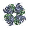
- PDB-5hnr: The X-ray structure of octameric human native 5-aminolaevulinic a... -
+ Open data

- Basic information
| Entry | Database: PDB / ID: 5hnr | |||||||||
|---|---|---|---|---|---|---|---|---|---|---|
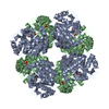
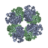
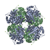
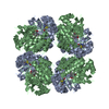
| Title | The X-ray structure of octameric human native 5-aminolaevulinic acid dehydratase. | |||||||||
 Components Components | Delta-aminolevulinic acid dehydratase | |||||||||
 Keywords Keywords | LYASE / Tetrapyrrole biosynthesis enzyme / TIM barrel / substrate complex. | |||||||||
| Function / homology |  Function and homology information Function and homology informationproteasome core complex binding / response to vitamin B1 / cellular response to lead ion / response to platinum ion / porphobilinogen synthase / porphobilinogen synthase activity / heme A biosynthetic process / response to aluminum ion / negative regulation of proteasomal protein catabolic process / heme B biosynthetic process ...proteasome core complex binding / response to vitamin B1 / cellular response to lead ion / response to platinum ion / porphobilinogen synthase / porphobilinogen synthase activity / heme A biosynthetic process / response to aluminum ion / negative regulation of proteasomal protein catabolic process / heme B biosynthetic process / response to mercury ion / : / response to selenium ion / response to cobalt ion / response to fatty acid / response to methylmercury / response to arsenic-containing substance / Heme biosynthesis / response to iron ion / heme biosynthetic process / response to herbicide / response to zinc ion / response to vitamin E / response to ionizing radiation / catalytic activity / response to amino acid / response to cadmium ion / cellular response to interleukin-4 / response to glucocorticoid / response to activity / protein homooligomerization / response to oxidative stress / secretory granule lumen / response to lipopolysaccharide / ficolin-1-rich granule lumen / response to ethanol / response to hypoxia / response to xenobiotic stimulus / Neutrophil degranulation / extracellular exosome / extracellular region / zinc ion binding / identical protein binding / nucleus / cytosol Similarity search - Function | |||||||||
| Biological species |  Homo sapiens (human) Homo sapiens (human) | |||||||||
| Method |  X-RAY DIFFRACTION / SYNCHROTRON / MOLECULAR REPLACEMENT / Resolution: 2.83 Å X-RAY DIFFRACTION / SYNCHROTRON / MOLECULAR REPLACEMENT / Resolution: 2.83 Å | |||||||||
 Authors Authors | Mills-Davies, N.L. / Thompson, D. / Shoolingin-Jordan, P.M. / Erskine, P.T. / Cooper, J.B. | |||||||||
 Citation Citation | Journal: Acta Crystallogr D Struct Biol / Year: 2017 Title: Structural studies of substrate and product complexes of 5-aminolaevulinic acid dehydratase from humans, Escherichia coli and the hyperthermophile Pyrobaculum calidifontis. Authors: Mills-Davies, N. / Butler, D. / Norton, E. / Thompson, D. / Sarwar, M. / Guo, J. / Gill, R. / Azim, N. / Coker, A. / Wood, S.P. / Erskine, P.T. / Coates, L. / Cooper, J.B. / Rashid, N. / ...Authors: Mills-Davies, N. / Butler, D. / Norton, E. / Thompson, D. / Sarwar, M. / Guo, J. / Gill, R. / Azim, N. / Coker, A. / Wood, S.P. / Erskine, P.T. / Coates, L. / Cooper, J.B. / Rashid, N. / Akhtar, M. / Shoolingin-Jordan, P.M. | |||||||||
| History |
|
- Structure visualization
| Structure viewer | Molecule: MolmilJmol/JSmol |
|---|
- Downloads & links
-Download
| PDBx/mmCIF format | 5hnr.cif.gz | 135.5 KB | Display | PDBx/mmCIF format |
|---|---|---|---|---|
| PDB format | pdb5hnr.ent.gz | 106 KB | Display | PDB format |
| PDBx/mmJSON format | 5hnr.json.gz | Tree view | PDBx/mmJSON format | |
| Others |  Other downloads Other downloads |
-Validation report
| Arichive directory | https://data.pdbj.org/pub/pdb/validation_reports/hn/5hnrftp://data.pdbj.org/pub/pdb/validation_reports/hn/5hnr | HTTPS FTP |
|---|
-Related structure data
| Related structure data |  5hmsC  5lzlC  5mhbC  1aw5S S: Starting model for refinement C: citing same article ( |
|---|---|
| Similar structure data |


-Links
 PDBj
PDBj


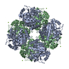
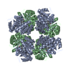
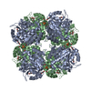
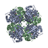
- Assembly
| Deposited unit | 
| ||||||||
|---|---|---|---|---|---|---|---|---|---|
| 1 | 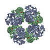
| ||||||||
| Unit cell |
|
-Components
| #1: Protein | Mass: 36338.852 Da / Num. of mol.: 2 / Source method: isolated from a natural source / Source: (natural) Homo sapiens (human) / Organ: Blood / References: UniProt: P13716, porphobilinogen synthase#2: Chemical | ChemComp-ZN / |   Mass: 65.409 Da / Num. of mol.: 1 / Source method: obtained synthetically / Formula: Zn Mass: 65.409 Da / Num. of mol.: 1 / Source method: obtained synthetically / Formula: Zn#3: Chemical |   Mass: 96.063 Da / Num. of mol.: 2 / Source method: obtained synthetically / Formula: SO4 Mass: 96.063 Da / Num. of mol.: 2 / Source method: obtained synthetically / Formula: SO4#4: Chemical | ChemComp-DAV / |   Mass: 118.154 Da / Num. of mol.: 1 / Source method: obtained synthetically / Formula: C5H12NO2 Mass: 118.154 Da / Num. of mol.: 1 / Source method: obtained synthetically / Formula: C5H12NO2#5: Water | ChemComp-HOH / |  Mass: 18.015 Da / Num. of mol.: 120 / Source method: isolated from a natural source / Formula: H2O Mass: 18.015 Da / Num. of mol.: 120 / Source method: isolated from a natural source / Formula: H2OHas protein modification | Y | |
|---|
-Experimental details
-Experiment
| Experiment | Method: X-RAY DIFFRACTION |
|---|
- Sample preparation
| Crystal | Density Matthews: 2.75 Å3/Da / Density % sol: 55.2 % |
|---|---|
| Crystal grow | Temperature: 293 K / Method: vapor diffusion, hanging drop / pH: 6.3 Details: 10 mg/ml protein concentration in 10 milliM Tris pH 7.4, 10 milliM diothiothreitol and 100 microM zinc chloride. 5 microlitres of this were mixed with an equal volume of 0.1 M MES pH range 6. ...Details: 10 mg/ml protein concentration in 10 milliM Tris pH 7.4, 10 milliM diothiothreitol and 100 microM zinc chloride. 5 microlitres of this were mixed with an equal volume of 0.1 M MES pH range 6.2 - 6.5, 1.0 - 1.6 M ammonium sulphate and 0 - 10 % dioxane. PH range: 6.2 - 6.5 |
-Data collection
| Diffraction | Mean temperature: 100 K |
|---|---|
| Diffraction source | Source: SYNCHROTRON / Site: SRS  / Beamline: PX9.5 / Wavelength: 0.87 Å / Beamline: PX9.5 / Wavelength: 0.87 Å |
| Detector | Type: MAR scanner 180 mm plate / Detector: IMAGE PLATE / Date: Jun 1, 1998 |
| Radiation | Protocol: SINGLE WAVELENGTH / Monochromatic (M) / Laue (L): M / Scattering type: x-ray |
| Radiation wavelength | Wavelength: 0.87 Å / Relative weight: 1 |
| Reflection | Resolution: 2.8→54.2 Å / Num. all: 18652 / Num. obs: 18652 / % possible obs: 96 % / Observed criterion σ(F): 0 / Observed criterion σ(I): 0 / Redundancy: 5.9 % / Biso Wilson estimate: 36.1 Å2 / Rmerge(I) obs: 0.071 / Net I/σ(I): 6 |
| Reflection shell | Resolution: 2.8→3 Å / Redundancy: 3.7 % / Rmerge(I) obs: 0.257 / Mean I/σ(I) obs: 2.9 / % possible all: 76.7 |
- Processing
| Software |
| ||||||||||||||||||||||||||||||||||||||||||||||||||||||||||||||||||||||||||||||||||||||||||||||||||||||||||||||||||||||||||||||||||||||||||||||||||||||||||||||||||||||||||||||||||||||
|---|---|---|---|---|---|---|---|---|---|---|---|---|---|---|---|---|---|---|---|---|---|---|---|---|---|---|---|---|---|---|---|---|---|---|---|---|---|---|---|---|---|---|---|---|---|---|---|---|---|---|---|---|---|---|---|---|---|---|---|---|---|---|---|---|---|---|---|---|---|---|---|---|---|---|---|---|---|---|---|---|---|---|---|---|---|---|---|---|---|---|---|---|---|---|---|---|---|---|---|---|---|---|---|---|---|---|---|---|---|---|---|---|---|---|---|---|---|---|---|---|---|---|---|---|---|---|---|---|---|---|---|---|---|---|---|---|---|---|---|---|---|---|---|---|---|---|---|---|---|---|---|---|---|---|---|---|---|---|---|---|---|---|---|---|---|---|---|---|---|---|---|---|---|---|---|---|---|---|---|---|---|---|---|
| Refinement | Method to determine structure: MOLECULAR REPLACEMENT Starting model: 1aw5 Resolution: 2.83→44.38 Å / Cor.coef. Fo:Fc: 0.957 / Cor.coef. Fo:Fc free: 0.906 / SU B: 13.612 / SU ML: 0.26 / Cross valid method: THROUGHOUT / ESU R Free: 0.367 / Stereochemistry target values: MAXIMUM LIKELIHOOD / Details: HYDROGENS HAVE BEEN ADDED IN THE RIDING POSITIONS
| ||||||||||||||||||||||||||||||||||||||||||||||||||||||||||||||||||||||||||||||||||||||||||||||||||||||||||||||||||||||||||||||||||||||||||||||||||||||||||||||||||||||||||||||||||||||
| Solvent computation | Ion probe radii: 0.8 Å / Shrinkage radii: 0.8 Å / VDW probe radii: 1.2 Å / Solvent model: MASK | ||||||||||||||||||||||||||||||||||||||||||||||||||||||||||||||||||||||||||||||||||||||||||||||||||||||||||||||||||||||||||||||||||||||||||||||||||||||||||||||||||||||||||||||||||||||
| Displacement parameters | Biso mean: 59.13 Å2
| ||||||||||||||||||||||||||||||||||||||||||||||||||||||||||||||||||||||||||||||||||||||||||||||||||||||||||||||||||||||||||||||||||||||||||||||||||||||||||||||||||||||||||||||||||||||
| Refinement step | Cycle: 1 / Resolution: 2.83→44.38 Å
| ||||||||||||||||||||||||||||||||||||||||||||||||||||||||||||||||||||||||||||||||||||||||||||||||||||||||||||||||||||||||||||||||||||||||||||||||||||||||||||||||||||||||||||||||||||||
| Refine LS restraints |
|