Mass: 18.015 Da / Num. of mol.: 327 / Source method: isolated from a natural source / Formula: H2O
-
Experimental details
-
Experiment
Experiment
Method: X-RAY DIFFRACTION / Number of used crystals: 1
-
Sample preparation
Crystal
Density Matthews: 4.14 Å3/Da / Density % sol: 70.3 %
Crystal grow
Temperature: 293 K / Method: vapor diffusion, hanging drop / pH: 8.2 Details: ALAD at 7 mg per ml mixed with equal volume of well solution: 200 mM Tris pH 8.0 - 8.4, 2 % saturated Ammonium sulphate, 200 microM zinc sulphate, 6 mM beta-mercaptoethanol and 3 mM ...Details: ALAD at 7 mg per ml mixed with equal volume of well solution: 200 mM Tris pH 8.0 - 8.4, 2 % saturated Ammonium sulphate, 200 microM zinc sulphate, 6 mM beta-mercaptoethanol and 3 mM porphobilinogen. Crystals were grown in the dark. PH range: 8.0 - 8.4 / Temp details: Room temperature.
-
Data collection
Diffraction
ID
Mean temperature (K)
Crystal-ID
1
100
1
2
100
1
Diffraction source
Source
Site
Beamline
ID
Wavelength (Å)
SYNCHROTRON
ESRF
BM14
1
1.069
SYNCHROTRON
EMBL/DESY, HAMBURG
X11
2
0.834
Detector
Type
ID
Detector
Date
MAR scanner 345 mm plate
1
IMAGE PLATE
Nov 6, 1997
MAR scanner 300 mm plate
2
IMAGE PLATE
Jun 3, 1998
Radiation
ID
Protocol
Monochromatic (M) / Laue (L)
Scattering type
Wavelength-ID
1
SINGLEWAVELENGTH
M
x-ray
1
2
SINGLEWAVELENGTH
M
x-ray
2
Radiation wavelength
ID
Wavelength (Å)
Relative weight
1
1.069
1
2
0.834
1
Reflection
Entry-ID: 5MHB
Resolution (Å)
Num. obs
% possible obs (%)
Redundancy (%)
Biso Wilson estimate (Å2)
CC1/2
Rmerge(I) obs
Diffraction-ID
Net I/σ(I)
2.1-36.8
34248
97.1
4.8
27.1
0.994
0.116
1
7.8
2.07-23.84
34487
95.2
7
0.998
0.078
2
15.3
Reflection shell
Resolution (Å)
Redundancy (%)
Rmerge(I) obs
Mean I/σ(I) obs
CC1/2
Diffraction-ID
% possible all
2.1-2.21
4.3
0.725
1.9
0.692
1
98
2.07-2.19
4.3
0.368
3.2
0.911
2
68.1
-
Processing
Software
Name
Version
Classification
MOSFLM
5.8.0107
datareduction
SCALA
datascaling
REFMAC
5.8.0107
refinement
Refinement
Method to determine structure: FOURIER SYNTHESIS Starting model: 1b4e
Resolution: 2.1→95.57 Å / Cor.coef. Fo:Fc: 0.967 / Cor.coef. Fo:Fc free: 0.938 / SU B: 7.475 / SU ML: 0.099 / Cross valid method: THROUGHOUT / ESU R: 0.123 / ESU R Free: 0.135 / Details: HYDROGENS HAVE BEEN ADDED IN THE RIDING POSITIONS
Rfactor
Num. reflection
% reflection
Selection details
Rfree
0.21285
1761
5.1 %
RANDOM
Rwork
0.15676
-
-
-
obs
0.15953
32987
98.95 %
-
Solvent computation
Ion probe radii: 0.8 Å / Shrinkage radii: 0.8 Å / VDW probe radii: 1.2 Å
 Movie
Movie Controller


 Open data
 Basic information
 Components
 Keywords
 Function and homology information
 X-RAY DIFFRACTION /
  Authors
 Citation
 Structure visualization
 Downloads & links
 Other downloads


 PDBj
PDBj
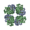
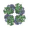
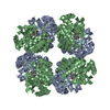
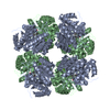
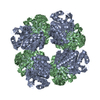
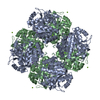
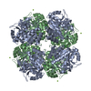
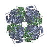
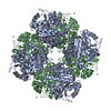
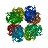
 Assembly
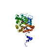

 Mass: 226.229 Da / Num. of mol.: 1 / Source method: obtained synthetically / Formula: C10H14N2O4
 Mass: 65.409 Da / Num. of mol.: 3 / Source method: obtained synthetically / Formula: Zn
 Mass: 92.094 Da / Num. of mol.: 3 / Source method: obtained synthetically / Formula: C3H8O3
 Mass: 18.015 Da / Num. of mol.: 327 / Source method: isolated from a natural source / Formula: H2O
 Sample preparation

 Processing