 Movie
Movie Controller

[English] 日本語
 Yorodumi
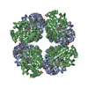
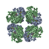
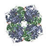
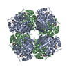
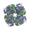
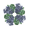
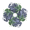
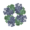
- PDB-1i8j: CRYSTAL STRUCTURE OF PORPHOBILINOGEN SYNTHASE COMPLEXED WITH THE ... -
+ Open data

- Basic information
| Entry | Database: PDB / ID: 1i8j | ||||||
|---|---|---|---|---|---|---|---|
| Title | CRYSTAL STRUCTURE OF PORPHOBILINOGEN SYNTHASE COMPLEXED WITH THE INHIBITOR 4,7-DIOXOSEBACIC ACID | ||||||
 Components Components | PORPHOBILINOGEN SYNTHASE | ||||||
 Keywords Keywords | LYASE / HEME BIOSYNTHESIS / MAGNESIUM / 4 / 7-DIOXOSEBACIC ACID | ||||||
| Function / homology |  Function and homology information Function and homology informationporphobilinogen synthase / porphobilinogen synthase activity / : / heme biosynthetic process / magnesium ion binding / zinc ion binding / cytosol Similarity search - Function | ||||||
| Biological species |  | ||||||
| Method |  X-RAY DIFFRACTION / MOLECULAR REPLACEMENT / Resolution: 1.9 Å X-RAY DIFFRACTION / MOLECULAR REPLACEMENT / Resolution: 1.9 Å | ||||||
 Authors Authors | Kervinen, J. / Jaffe, E.K. / Stauffer, F. / Neier, R. / Wlodawer, A. / Zdanov, A. | ||||||
 Citation Citation | Journal: Biochemistry / Year: 2001 Title: Mechanistic basis for suicide inactivation of porphobilinogen synthase by 4,7-dioxosebacic acid, an inhibitor that shows dramatic species selectivity. Authors: Kervinen, J. / Jaffe, E.K. / Stauffer, F. / Neier, R. / Wlodawer, A. / Zdanov, A. | ||||||
| History |
| ||||||
| Remark 600 | HETEROGEN Atoms C4 and C7 of DSB form a Schiff base to NZ of Lys246 and NZ of Lys194. The oxygens ...HETEROGEN Atoms C4 and C7 of DSB form a Schiff base to NZ of Lys246 and NZ of Lys194. The oxygens on the C4 and C7 are eliminated during this reaction. |
- Structure visualization
| Structure viewer | Molecule: MolmilJmol/JSmol |
|---|
- Downloads & links
-Download
| PDBx/mmCIF format | 1i8j.cif.gz | 141.5 KB | Display | PDBx/mmCIF format |
|---|---|---|---|---|
| PDB format | pdb1i8j.ent.gz | 111.4 KB | Display | PDB format |
| PDBx/mmJSON format | 1i8j.json.gz | Tree view | PDBx/mmJSON format | |
| Others |  Other downloads Other downloads |
-Validation report
| Arichive directory | https://data.pdbj.org/pub/pdb/validation_reports/i8/1i8jftp://data.pdbj.org/pub/pdb/validation_reports/i8/1i8j | HTTPS FTP |
|---|
-Related structure data
| Related structure data | 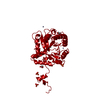 1b4eS S: Starting model for refinement |
|---|---|
| Similar structure data |

-Links
 PDBj
PDBj
- Assembly
| Deposited unit | 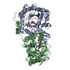
| ||||||||
|---|---|---|---|---|---|---|---|---|---|
| 1 | 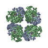
| ||||||||
| Unit cell |
|
-Components
| #1: Protein | Mass: 35537.609 Da / Num. of mol.: 2 Source method: isolated from a genetically manipulated source Source: (gene. exp.) #2: Chemical |   Mass: 65.409 Da / Num. of mol.: 2 / Source method: obtained synthetically / Formula: Zn Mass: 65.409 Da / Num. of mol.: 2 / Source method: obtained synthetically / Formula: Zn#3: Chemical |   Mass: 24.305 Da / Num. of mol.: 2 / Source method: obtained synthetically / Formula: Mg Mass: 24.305 Da / Num. of mol.: 2 / Source method: obtained synthetically / Formula: Mg#4: Chemical | 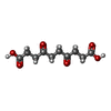  Mass: 230.215 Da / Num. of mol.: 2 / Source method: obtained synthetically / Formula: C10H14O6 Mass: 230.215 Da / Num. of mol.: 2 / Source method: obtained synthetically / Formula: C10H14O6#5: Water | ChemComp-HOH / |  Mass: 18.015 Da / Num. of mol.: 316 / Source method: isolated from a natural source / Formula: H2O Mass: 18.015 Da / Num. of mol.: 316 / Source method: isolated from a natural source / Formula: H2OHas protein modification | Y | Nonpolymer details | ATOMS C4 AND C7 OF DSB FORM A SCHIFF BASE TO NZ OF LYS246 AND NZ OF LYS194. THE OXYGENS ON THE C4 ...ATOMS C4 AND C7 OF DSB FORM A SCHIFF BASE TO NZ OF LYS246 AND NZ OF LYS194. THE OXYGENS ON THE C4 AND C7 ARE ELIMINATED | |
|---|
-Experimental details
-Experiment
| Experiment | Method: X-RAY DIFFRACTION / Number of used crystals: 1 |
|---|
- Sample preparation
| Crystal | Density Matthews: 4.16 Å3/Da / Density % sol: 70.43 % | ||||||||||||||||||||||||||||||||||||||||||||||||||||||||||||
|---|---|---|---|---|---|---|---|---|---|---|---|---|---|---|---|---|---|---|---|---|---|---|---|---|---|---|---|---|---|---|---|---|---|---|---|---|---|---|---|---|---|---|---|---|---|---|---|---|---|---|---|---|---|---|---|---|---|---|---|---|---|
| Crystal grow | Temperature: 294 K / Method: vapor diffusion, sitting drop / pH: 8.5 Details: PEG 3350, GLYCEROL, TRIS-HCL, pH 8.5, VAPOR DIFFUSION, SITTING DROP, temperature 294K | ||||||||||||||||||||||||||||||||||||||||||||||||||||||||||||
| Crystal grow | *PLUS pH: 8 | ||||||||||||||||||||||||||||||||||||||||||||||||||||||||||||
| Components of the solutions | *PLUS
|
-Data collection
| Diffraction | Mean temperature: 100 K |
|---|---|
| Diffraction source | Source: ROTATING ANODE / Type: RIGAKU RU200 / Wavelength: 1.5418 Å |
| Detector | Type: MARRESEARCH / Detector: IMAGE PLATE / Date: Nov 10, 2000 / Details: OSMIC |
| Radiation | Protocol: SINGLE WAVELENGTH / Monochromatic (M) / Laue (L): M / Scattering type: x-ray |
| Radiation wavelength | Wavelength: 1.5418 Å / Relative weight: 1 |
| Reflection | Resolution: 1.9→35 Å / Num. all: 94638 / Num. obs: 94536 / % possible obs: 99.9 % / Observed criterion σ(F): 2 / Observed criterion σ(I): 1 / Redundancy: 5.6 % / Rmerge(I) obs: 0.069 / Net I/σ(I): 12 |
| Reflection shell | Resolution: 1.9→1.93 Å / Redundancy: 3.5 % / Rmerge(I) obs: 0.5 / % possible all: 99.8 |
| Reflection | *PLUS Lowest resolution: 35 Å |
- Processing
| Software |
| ||||||||||||||||||||
|---|---|---|---|---|---|---|---|---|---|---|---|---|---|---|---|---|---|---|---|---|---|
| Refinement | Method to determine structure: MOLECULAR REPLACEMENT Starting model: 1b4e Resolution: 1.9→35 Å / σ(F): 0 / σ(I): 0 / Stereochemistry target values: Engh & Huber
| ||||||||||||||||||||
| Refinement step | Cycle: LAST / Resolution: 1.9→35 Å
| ||||||||||||||||||||
| Refine LS restraints |
| ||||||||||||||||||||
| Software | *PLUS Name: CNS / Classification: refinement | ||||||||||||||||||||
| Refinement | *PLUS Highest resolution: 1.9 Å / Lowest resolution: 35 Å / σ(F): 0 / Rfactor obs: 0.198 | ||||||||||||||||||||
| Solvent computation | *PLUS | ||||||||||||||||||||
| Displacement parameters | *PLUS |
