 Movie
Movie Controller

[English] 日本語
 Yorodumi
- PDB-1b4e: X-ray structure of 5-aminolevulinic acid dehydratase complexed wi... -
+ Open data

- Basic information
| Entry | Database: PDB / ID: 1b4e | ||||||
|---|---|---|---|---|---|---|---|
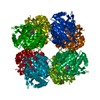
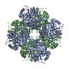
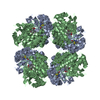
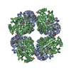
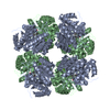
| Title | X-ray structure of 5-aminolevulinic acid dehydratase complexed with the inhibitor levulinic acid | ||||||
 Components Components | PROTEIN (5-AMINOLEVULINIC ACID DEHYDRATASE) | ||||||
 Keywords Keywords | LYASE / DEHYDRATASE | ||||||
| Function / homology |  Function and homology information Function and homology informationporphobilinogen synthase / porphobilinogen synthase activity / : / heme biosynthetic process / magnesium ion binding / zinc ion binding / cytosol Similarity search - Function | ||||||
| Biological species |  | ||||||
| Method |  X-RAY DIFFRACTION / SYNCHROTRON / MOLECULAR REPLACEMENT / Resolution: 2 Å X-RAY DIFFRACTION / SYNCHROTRON / MOLECULAR REPLACEMENT / Resolution: 2 Å | ||||||
 Authors Authors | Erskine, P.T. / Cooper, J.B. / Lewis, G. / Spencer, P. / Wood, S.P. / Shoolingin-Jordan, P.M. | ||||||
 Citation Citation | Journal: Biochemistry / Year: 1999 Title: X-ray structure of 5-aminolevulinic acid dehydratase from Escherichia coli complexed with the inhibitor levulinic acid at 2.0 A resolution. Authors: Erskine, P.T. / Norton, E. / Cooper, J.B. / Lambert, R. / Coker, A. / Lewis, G. / Spencer, P. / Sarwar, M. / Wood, S.P. / Warren, M.J. / Shoolingin-Jordan, P.M. | ||||||
| History |
|
- Structure visualization
| Structure viewer | Molecule: MolmilJmol/JSmol |
|---|
- Downloads & links
-Download
| PDBx/mmCIF format | 1b4e.cif.gz | 86.5 KB | Display | PDBx/mmCIF format |
|---|---|---|---|---|
| PDB format | pdb1b4e.ent.gz | 63.6 KB | Display | PDB format |
| PDBx/mmJSON format | 1b4e.json.gz | Tree view | PDBx/mmJSON format | |
| Others |  Other downloads Other downloads |
-Validation report
| Arichive directory | https://data.pdbj.org/pub/pdb/validation_reports/b4/1b4eftp://data.pdbj.org/pub/pdb/validation_reports/b4/1b4e | HTTPS FTP |
|---|
-Related structure data
| Similar structure data |
|---|

-Links
 PDBj
PDBj
- Assembly
| Deposited unit | 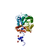
| ||||||||
|---|---|---|---|---|---|---|---|---|---|
| 1 | x 8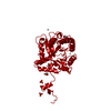
| ||||||||
| Unit cell |
|
-Components
-Protein , 1 types, 1 molecules A
| #1: Protein | Mass: 35537.566 Da / Num. of mol.: 1 Source method: isolated from a genetically manipulated source Source: (gene. exp.) |
|---|
-Non-polymers , 5 types, 381 molecules 


| #2: Chemical |  Mass: 96.063 Da / Num. of mol.: 3 / Source method: obtained synthetically / Formula: SO4 Mass: 96.063 Da / Num. of mol.: 3 / Source method: obtained synthetically / Formula: SO4#3: Chemical |  Mass: 65.409 Da / Num. of mol.: 3 / Source method: obtained synthetically / Formula: Zn Mass: 65.409 Da / Num. of mol.: 3 / Source method: obtained synthetically / Formula: Zn#4: Chemical | ChemComp-SHF / |  Mass: 116.115 Da / Num. of mol.: 1 / Source method: obtained synthetically / Formula: C5H8O3 Mass: 116.115 Da / Num. of mol.: 1 / Source method: obtained synthetically / Formula: C5H8O3#5: Chemical |  Mass: 92.094 Da / Num. of mol.: 3 / Source method: obtained synthetically / Formula: C3H8O3 Mass: 92.094 Da / Num. of mol.: 3 / Source method: obtained synthetically / Formula: C3H8O3#6: Water | ChemComp-HOH / | Mass: 18.015 Da / Num. of mol.: 371 / Source method: isolated from a natural source / Formula: H2O |
|---|
-Details
| Has protein modification | Y |
|---|
-Experimental details
-Experiment
| Experiment | Method: X-RAY DIFFRACTION / Number of used crystals: 1 |
|---|
- Sample preparation
| Crystal | Density Matthews: 4 Å3/Da / Density % sol: 71 % | |||||||||||||||||||||||||||||||||||
|---|---|---|---|---|---|---|---|---|---|---|---|---|---|---|---|---|---|---|---|---|---|---|---|---|---|---|---|---|---|---|---|---|---|---|---|---|
| Crystal grow | Method: vapor diffusion, hanging drop / pH: 8.2 Details: 7MG/ML ENZYME AND 2 - 5 % SATURATED AMMONIUM SULPHATE AS PRECIPITANT. 0.2 M TRIS-HCL USED TO BUFFER THE PH BETWEEN 8.1 AND 8.4. 15MM LEVULINIC ACID, 40UM ZINC SULPHATE AND 4MM BETA- ...Details: 7MG/ML ENZYME AND 2 - 5 % SATURATED AMMONIUM SULPHATE AS PRECIPITANT. 0.2 M TRIS-HCL USED TO BUFFER THE PH BETWEEN 8.1 AND 8.4. 15MM LEVULINIC ACID, 40UM ZINC SULPHATE AND 4MM BETA- MERCAPTOETHANOL WERE PRESENT., pH 8.2, VAPOR DIFFUSION, HANGING DROP | |||||||||||||||||||||||||||||||||||
| Crystal grow | *PLUS PH range low: 8.4 / PH range high: 8.1 | |||||||||||||||||||||||||||||||||||
| Components of the solutions | *PLUS
|
-Data collection
| Diffraction | Mean temperature: 100 K |
|---|---|
| Diffraction source | Source: SYNCHROTRON / Site: SRS  / Beamline: PX9.6 / Wavelength: 0.87 / Beamline: PX9.6 / Wavelength: 0.87 |
| Detector | Type: MAR scanner 300 mm plate / Detector: IMAGE PLATE / Date: Mar 15, 1994 |
| Radiation | Protocol: SINGLE WAVELENGTH / Monochromatic (M) / Laue (L): M / Scattering type: x-ray |
| Radiation wavelength | Wavelength: 0.87 Å / Relative weight: 1 |
| Reflection | Resolution: 2→8 Å / Num. obs: 33275 / % possible obs: 85.8 % / Observed criterion σ(I): 0 / Redundancy: 2.5 % / Rmerge(I) obs: 0.063 / Net I/σ(I): 8.8 |
| Reflection shell | Resolution: 2→2.1 Å / Redundancy: 1.6 % / Rmerge(I) obs: 0.297 / Mean I/σ(I) obs: 2.1 / % possible all: 70.8 |
| Reflection | *PLUS Lowest resolution: 7.7 Å |
| Reflection shell | *PLUS % possible obs: 70.8 % |
- Processing
| Software |
| ||||||||||||||||||||||||||||||||||||||||||||||||||||||||||||
|---|---|---|---|---|---|---|---|---|---|---|---|---|---|---|---|---|---|---|---|---|---|---|---|---|---|---|---|---|---|---|---|---|---|---|---|---|---|---|---|---|---|---|---|---|---|---|---|---|---|---|---|---|---|---|---|---|---|---|---|---|---|
| Refinement | Method to determine structure: MOLECULAR REPLACEMENT Starting model: YEAST ALAD Resolution: 2→8 Å / Cross valid method: THROUGHOUT / σ(F): 0
| ||||||||||||||||||||||||||||||||||||||||||||||||||||||||||||
| Refinement step | Cycle: LAST / Resolution: 2→8 Å
| ||||||||||||||||||||||||||||||||||||||||||||||||||||||||||||
| Refine LS restraints |
| ||||||||||||||||||||||||||||||||||||||||||||||||||||||||||||
| Software | *PLUS Name: RESTRAIN / Classification: refinement | ||||||||||||||||||||||||||||||||||||||||||||||||||||||||||||
| Refinement | *PLUS Lowest resolution: 7.7 Å | ||||||||||||||||||||||||||||||||||||||||||||||||||||||||||||
| Solvent computation | *PLUS | ||||||||||||||||||||||||||||||||||||||||||||||||||||||||||||
| Displacement parameters | *PLUS |
