 Movie
Movie Controller

[English] 日本語
 Yorodumi
- PDB-1gzg: Complex of a Mg2-dependent porphobilinogen synthase from Pseudomo... -
+ Open data

- Basic information
| Entry | Database: PDB / ID: 1gzg | |||||||||
|---|---|---|---|---|---|---|---|---|---|---|
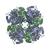
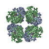
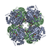
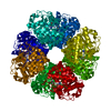
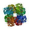
| Title | Complex of a Mg2-dependent porphobilinogen synthase from Pseudomonas aeruginosa (mutant D139N) with 5-fluorolevulinic acid | |||||||||
 Components Components | DELTA-AMINOLEVULINIC ACID DEHYDRATASE | |||||||||
 Keywords Keywords | LYASE / HEME BIOSYNTHESIS / 5-FLUOROLEVULINIC ACID | |||||||||
| Function / homology |  Function and homology information Function and homology informationporphobilinogen synthase / porphobilinogen synthase activity / porphyrin-containing compound biosynthetic process / : / heme biosynthetic process / zinc ion binding / cytosol Similarity search - Function | |||||||||
| Biological species |   PSEUDOMONAS AERUGINOSA (bacteria) PSEUDOMONAS AERUGINOSA (bacteria) | |||||||||
| Method |  X-RAY DIFFRACTION / SYNCHROTRON / MOLECULAR REPLACEMENT / Resolution: 1.66 Å X-RAY DIFFRACTION / SYNCHROTRON / MOLECULAR REPLACEMENT / Resolution: 1.66 Å | |||||||||
 Authors Authors | Frere, F. / Schubert, W.-D. / Stauffer, F. / Frankenberg, N. / Neier, R. / Jahn, D. / Heinz, D.W. | |||||||||
 Citation Citation | Journal: J. Mol. Biol. / Year: 2002 Title: Structure of porphobilinogen synthase from Pseudomonas aeruginosa in complex with 5-fluorolevulinic acid suggests a double Schiff base mechanism. Authors: Frere, F. / Schubert, W.D. / Stauffer, F. / Frankenberg, N. / Neier, R. / Jahn, D. / Heinz, D.W. | |||||||||
| History |
| |||||||||
| Remark 700 | SHEET DETERMINATION METHOD: DSSP THE SHEETS PRESENTED AS "BA" IN EACH CHAIN ON SHEET RECORDS BELOW ... SHEET DETERMINATION METHOD: DSSP THE SHEETS PRESENTED AS "BA" IN EACH CHAIN ON SHEET RECORDS BELOW IS ACTUALLY AN 11-STRANDED BARREL THIS IS REPRESENTED BY A 12-STRANDED SHEET IN WHICH THE FIRST AND LAST STRANDS ARE IDENTICAL. |
- Structure visualization
| Structure viewer | Molecule: MolmilJmol/JSmol |
|---|
- Downloads & links
-Download
| PDBx/mmCIF format | 1gzg.cif.gz | 162.4 KB | Display | PDBx/mmCIF format |
|---|---|---|---|---|
| PDB format | pdb1gzg.ent.gz | 129.2 KB | Display | PDB format |
| PDBx/mmJSON format | 1gzg.json.gz | Tree view | PDBx/mmJSON format | |
| Others |  Other downloads Other downloads |
-Validation report
| Arichive directory | https://data.pdbj.org/pub/pdb/validation_reports/gz/1gzgftp://data.pdbj.org/pub/pdb/validation_reports/gz/1gzg | HTTPS FTP |
|---|
-Related structure data
| Related structure data | 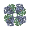 1b4kS S: Starting model for refinement |
|---|---|
| Similar structure data |

-Links
 PDBj
PDBj
- Assembly
| Deposited unit | 
| ||||||||||||
|---|---|---|---|---|---|---|---|---|---|---|---|---|---|
| 1 | 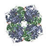
| ||||||||||||
| Unit cell |
| ||||||||||||
| Components on special symmetry positions |
|
-Components
-Protein , 1 types, 2 molecules AB
| #1: Protein | Mass: 37062.914 Da / Num. of mol.: 2 / Mutation: YES Source method: isolated from a genetically manipulated source Details: SCHIFF BASE LINKS BETWEEN ATOMS NZ OF LYS205 AND LYS260 AND ATOMS C5 OF 5-FLUOROLEVULINIC ACID Source: (gene. exp.) PSEUDOMONAS AERUGINOSA (bacteria) / Plasmid: PGEX-6P-1 / Production host: |
|---|
-Non-polymers , 6 types, 683 molecules 


| #2: Chemical | ChemComp-LAF /  Mass: 134.106 Da / Num. of mol.: 4 / Source method: obtained synthetically / Formula: C5H7FO3 Mass: 134.106 Da / Num. of mol.: 4 / Source method: obtained synthetically / Formula: C5H7FO3#3: Chemical |  Mass: 24.305 Da / Num. of mol.: 2 / Source method: obtained synthetically / Formula: Mg Mass: 24.305 Da / Num. of mol.: 2 / Source method: obtained synthetically / Formula: Mg#4: Chemical |  Mass: 22.990 Da / Num. of mol.: 2 / Source method: obtained synthetically / Formula: Na Mass: 22.990 Da / Num. of mol.: 2 / Source method: obtained synthetically / Formula: Na#5: Chemical |  Mass: 96.063 Da / Num. of mol.: 2 / Source method: obtained synthetically / Formula: SO4 Mass: 96.063 Da / Num. of mol.: 2 / Source method: obtained synthetically / Formula: SO4#6: Chemical |  Mass: 39.098 Da / Num. of mol.: 2 / Source method: obtained synthetically / Formula: K Mass: 39.098 Da / Num. of mol.: 2 / Source method: obtained synthetically / Formula: K#7: Water | ChemComp-HOH / | Mass: 18.015 Da / Num. of mol.: 671 / Source method: isolated from a natural source / Formula: H2O |
|---|
-Details
| Has protein modification | Y |
|---|---|
| Sequence details | MUTANT D139N, THE CRYSTALLIZ |
-Experimental details
-Experiment
| Experiment | Method: X-RAY DIFFRACTION / Number of used crystals: 1 |
|---|
- Sample preparation
| Crystal | Density Matthews: 2.46 Å3/Da / Density % sol: 50 % | ||||||||||||||||||||||||||||||||||||||||||
|---|---|---|---|---|---|---|---|---|---|---|---|---|---|---|---|---|---|---|---|---|---|---|---|---|---|---|---|---|---|---|---|---|---|---|---|---|---|---|---|---|---|---|---|
| Crystal grow | Method: vapor diffusion, sitting drop / pH: 9 Details: PROTEIN SOLUTION: 12 MG/ML PBGS MUTANT D139N IN 50 MM K-HEPES, 10 MM MGCL2. CRYSTALLIZATION SOLUTION: 0.99 ML PROTEIN SOLUTION = 0.01 ML 2MM 5F-LA IN 50 MM K-HEPES, 10 MM MGCL2. RESERVOIR ...Details: PROTEIN SOLUTION: 12 MG/ML PBGS MUTANT D139N IN 50 MM K-HEPES, 10 MM MGCL2. CRYSTALLIZATION SOLUTION: 0.99 ML PROTEIN SOLUTION = 0.01 ML 2MM 5F-LA IN 50 MM K-HEPES, 10 MM MGCL2. RESERVOIR SOLUTION: 1 M NA/K-TARTRATE, 0.2M LI2SO4, 0.1 M CHES PH = 9,5. SITTING DROP: 0.003 ML CRYSTALLISATION SOLUTION + 0.003 ML RESERVOIR SOLUTION | ||||||||||||||||||||||||||||||||||||||||||
| Crystal grow | *PLUS pH: 7.5 / Method: sparse matrix screening | ||||||||||||||||||||||||||||||||||||||||||
| Components of the solutions | *PLUS
|
-Data collection
| Diffraction | Mean temperature: 100 K |
|---|---|
| Diffraction source | Source: SYNCHROTRON / Site: MPG/DESY, HAMBURG  / Beamline: BW6 / Wavelength: 1.05 / Beamline: BW6 / Wavelength: 1.05 |
| Detector | Type: MARRESEARCH / Detector: IMAGE PLATE / Date: Oct 15, 2001 / Details: MIRRORS |
| Radiation | Protocol: SINGLE WAVELENGTH / Monochromatic (M) / Laue (L): M / Scattering type: x-ray |
| Radiation wavelength | Wavelength: 1.05 Å / Relative weight: 1 |
| Reflection | Resolution: 1.66→20 Å / Num. obs: 82775 / % possible obs: 99.9 % / Observed criterion σ(I): 3 / Redundancy: 5.2 % / Rmerge(I) obs: 0.116 / Net I/σ(I): 11 |
| Reflection shell | Resolution: 1.66→1.72 Å / Redundancy: 4.9 % / Rmerge(I) obs: 0.394 / Mean I/σ(I) obs: 4.1 / % possible all: 100 |
| Reflection | *PLUS Highest resolution: 1.66 Å / Lowest resolution: 20 Å / Num. measured all: 433947 |
| Reflection shell | *PLUS % possible obs: 100 % / Redundancy: 4.9 % / Mean I/σ(I) obs: 4.1 |
- Processing
| Software |
| ||||||||||||||||||||||||||||||||||||||||||||||||||||||||||||||||||||||||||||||||||||||||||||||||||||||||||||||||||||||||||||||||||||||||||||||||||||||||||||||||||||||||||||||||||||||
|---|---|---|---|---|---|---|---|---|---|---|---|---|---|---|---|---|---|---|---|---|---|---|---|---|---|---|---|---|---|---|---|---|---|---|---|---|---|---|---|---|---|---|---|---|---|---|---|---|---|---|---|---|---|---|---|---|---|---|---|---|---|---|---|---|---|---|---|---|---|---|---|---|---|---|---|---|---|---|---|---|---|---|---|---|---|---|---|---|---|---|---|---|---|---|---|---|---|---|---|---|---|---|---|---|---|---|---|---|---|---|---|---|---|---|---|---|---|---|---|---|---|---|---|---|---|---|---|---|---|---|---|---|---|---|---|---|---|---|---|---|---|---|---|---|---|---|---|---|---|---|---|---|---|---|---|---|---|---|---|---|---|---|---|---|---|---|---|---|---|---|---|---|---|---|---|---|---|---|---|---|---|---|---|
| Refinement | Method to determine structure: MOLECULAR REPLACEMENT Starting model: 1B4K Resolution: 1.66→91.29 Å / Cor.coef. Fo:Fc: 0.967 / Cor.coef. Fo:Fc free: 0.954 / SU B: 2.31 / SU ML: 0.08 / Cross valid method: THROUGHOUT / ESU R: 0.099 / ESU R Free: 0.094 / Stereochemistry target values: MAXIMUM LIKELIHOOD Details: LAST TWO C-TERMINAL RESIDUES WERE NOT SEEN IN THE DENSITY MAP
| ||||||||||||||||||||||||||||||||||||||||||||||||||||||||||||||||||||||||||||||||||||||||||||||||||||||||||||||||||||||||||||||||||||||||||||||||||||||||||||||||||||||||||||||||||||||
| Solvent computation | Ion probe radii: 0.8 Å / Shrinkage radii: 0.8 Å / VDW probe radii: 1.4 Å / Solvent model: BABINET MODEL PLUS MASK | ||||||||||||||||||||||||||||||||||||||||||||||||||||||||||||||||||||||||||||||||||||||||||||||||||||||||||||||||||||||||||||||||||||||||||||||||||||||||||||||||||||||||||||||||||||||
| Displacement parameters | Biso mean: 20.86 Å2
| ||||||||||||||||||||||||||||||||||||||||||||||||||||||||||||||||||||||||||||||||||||||||||||||||||||||||||||||||||||||||||||||||||||||||||||||||||||||||||||||||||||||||||||||||||||||
| Refinement step | Cycle: LAST / Resolution: 1.66→91.29 Å
| ||||||||||||||||||||||||||||||||||||||||||||||||||||||||||||||||||||||||||||||||||||||||||||||||||||||||||||||||||||||||||||||||||||||||||||||||||||||||||||||||||||||||||||||||||||||
| Refine LS restraints |
|