 Movie
Movie Controller

[English] 日本語
 Yorodumi
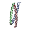
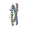
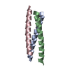
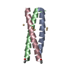
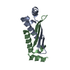
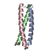
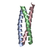
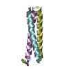
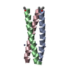
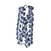
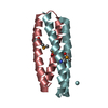
- PDB-3k7z: GCN4-Leucine zipper core mutant as N16A trigonal automatic solution -
+ Open data

- Basic information
| Entry | Database: PDB / ID: 3k7z | |||||||||
|---|---|---|---|---|---|---|---|---|---|---|
| Title | GCN4-Leucine zipper core mutant as N16A trigonal automatic solution | |||||||||
 Components Components | General control protein GCN4 | |||||||||
 Keywords Keywords | DNA BINDING PROTEIN / COILED COIL / LEUCINE ZIPPER / Structural Genomics / TB Structural Genomics Consortium / TBSGC / Activator / Amino-acid biosynthesis / DNA-binding / Nucleus / Phosphoprotein / Transcription / Transcription regulation | |||||||||
| Function / homology |  Function and homology information Function and homology informationFCERI mediated MAPK activation / protein localization to nuclear periphery / Activation of the AP-1 family of transcription factors / negative regulation of ribosomal protein gene transcription by RNA polymerase II / positive regulation of cellular response to amino acid starvation / response to amino acid starvation / mediator complex binding / Oxidative Stress Induced Senescence / amino acid biosynthetic process / TFIID-class transcription factor complex binding ...FCERI mediated MAPK activation / protein localization to nuclear periphery / Activation of the AP-1 family of transcription factors / negative regulation of ribosomal protein gene transcription by RNA polymerase II / positive regulation of cellular response to amino acid starvation / response to amino acid starvation / mediator complex binding / Oxidative Stress Induced Senescence / amino acid biosynthetic process / TFIID-class transcription factor complex binding / positive regulation of RNA polymerase II transcription preinitiation complex assembly / positive regulation of transcription initiation by RNA polymerase II / cellular response to nutrient levels / cellular response to amino acid starvation / RNA polymerase II transcription regulator complex / DNA-binding transcription activator activity, RNA polymerase II-specific / transcription regulator complex / sequence-specific DNA binding / RNA polymerase II-specific DNA-binding transcription factor binding / DNA-binding transcription factor activity, RNA polymerase II-specific / intracellular signal transduction / RNA polymerase II cis-regulatory region sequence-specific DNA binding / DNA-binding transcription factor activity / chromatin binding / negative regulation of transcription by RNA polymerase II / positive regulation of transcription by RNA polymerase II / identical protein binding / nucleus Similarity search - Function | |||||||||
| Method |  X-RAY DIFFRACTION / SYNCHROTRON / MAD / Resolution: 1.9 Å X-RAY DIFFRACTION / SYNCHROTRON / MAD / Resolution: 1.9 Å | |||||||||
 Authors Authors | Holton, J. / Alber, T. / TB Structural Genomics Consortium (TBSGC) | |||||||||
 Citation Citation | Journal: Proc.Natl.Acad.Sci.USA / Year: 2004 Title: Automated Protein Crystal Structure Determination Using Elves. Authors: Holton, J. / Alber, T. #1: Journal: Nat.Struct.Biol. / Year: 1996Title: An Engineered Allosteric Switch in Leucine-Zipper Oligomerization Authors: Gonzalez Jr., L. / Plecs, J.J. / Alber, T. #2: Journal: Nature / Year: 1994Title: Crystal Structure of an Isoleucine-Zipper Trimer Authors: Harbury, P.B. / Kim, P.S. / Alber, T. #3: Journal: Science / Year: 1993Title: A Switch between Two-, Three-, and Four-Stranded Coiled Coils in GCN4 Leucine Zipper Mutants Authors: Harbury, P.B. / Zhang, T. / Kim, P.S. / Alber, T. #4: Journal: Science / Year: 1991Title: X-Ray Structure of the GCN4 Leucine Zipper, a Two-Stranded, Parallel Coiled Coil. Authors: O'Shea, E.K. / Klemm, J.D. / Kim, P.S. / Alber, T. | |||||||||
| History |
|
- Structure visualization
| Structure viewer | Molecule: MolmilJmol/JSmol |
|---|
- Downloads & links
-Download
| PDBx/mmCIF format | 3k7z.cif.gz | 52 KB | Display | PDBx/mmCIF format |
|---|---|---|---|---|
| PDB format | pdb3k7z.ent.gz | 32.9 KB | Display | PDB format |
| PDBx/mmJSON format | 3k7z.json.gz | Tree view | PDBx/mmJSON format | |
| Others |  Other downloads Other downloads |
-Validation report
| Arichive directory | https://data.pdbj.org/pub/pdb/validation_reports/k7/3k7zftp://data.pdbj.org/pub/pdb/validation_reports/k7/3k7z | HTTPS FTP |
|---|
-Related structure data


-Links
 PDBj
PDBj

- Assembly
| Deposited unit | 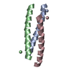
| ||||||||
|---|---|---|---|---|---|---|---|---|---|
| 1 |
| ||||||||
| Unit cell |
|
-Components
| #1: Protein/peptide | Mass: 3962.639 Da / Num. of mol.: 3 / Fragment: LEUCINE-ZIPPER (RESIDUES 249-281) / Mutation: N16A / Source method: obtained synthetically Details: THE SEQUENCE OF THE PROTEIN IS NATURALLY FOUND IN SACCHAROMYCES CEREVISIAE. THE PROTEIN WAS SYNTHESIZED BY SOLID PHASE PEPTIDE SYNTHESIS. References: UniProt: P03069 #2: Water | ChemComp-HOH / |  Mass: 18.015 Da / Num. of mol.: 621 / Source method: isolated from a natural source / Formula: H2O Mass: 18.015 Da / Num. of mol.: 621 / Source method: isolated from a natural source / Formula: H2O |
|---|
-Experimental details
-Experiment
| Experiment | Method: X-RAY DIFFRACTION / Number of used crystals: 1 |
|---|
- Sample preparation
| Crystal | Density Matthews: 4.54 Å3/Da / Density % sol: 75 % |
|---|---|
| Crystal grow | Temperature: 298 K / Method: vapor diffusion, sitting drop / pH: 7.3 Details: 100MM BIS-TRIS buffer only, pH 7.3, VAPOR DIFFUSION, SITTING DROP, temperature 298K |
-Data collection
| Diffraction | Mean temperature: 90 K | |||||||||||||||
|---|---|---|---|---|---|---|---|---|---|---|---|---|---|---|---|---|
| Diffraction source | Source: SYNCHROTRON / Site: SSRL  / Beamline: BL1-5 / Wavelength: 1.0688, 0.9800, 0.9795, 0.9322 / Beamline: BL1-5 / Wavelength: 1.0688, 0.9800, 0.9795, 0.9322 | |||||||||||||||
| Detector | Type: ADSC QUANTUM 4 / Detector: CCD / Date: Dec 5, 1997 | |||||||||||||||
| Radiation | Monochromator: SI(111) / Protocol: MAD / Monochromatic (M) / Laue (L): M / Scattering type: x-ray | |||||||||||||||
| Radiation wavelength |
| |||||||||||||||
| Reflection | Resolution: 1.8→45 Å / Num. all: 23719 / Num. obs: 23195 / % possible obs: 99.6 % / Observed criterion σ(F): 0 / Observed criterion σ(I): 0 / Redundancy: 11 % / Rmerge(I) obs: 0.097 | |||||||||||||||
| Reflection shell | Resolution: 1.8→1.9 Å / % possible all: 98.4 |
- Processing
| Software |
| |||||||||||||||||||||||||
|---|---|---|---|---|---|---|---|---|---|---|---|---|---|---|---|---|---|---|---|---|---|---|---|---|---|---|
| Refinement | Method to determine structure: MAD / Resolution: 1.9→25.96 Å / Cross valid method: THROUGHOUT / σ(F): 0 / Stereochemistry target values: refmac Details: THIS ENTRY WAS SOLVED USING AUTOMATED STRUCTURE WHILE RELATED ENTRTY 1RB5 WAS SOLVED USING NON-AUTOMATED STRUCTURE DETERMINATION METHOD. THE NUMBER OF CLOSE CONTACTS IN THIS STRUCTURE IS AN ...Details: THIS ENTRY WAS SOLVED USING AUTOMATED STRUCTURE WHILE RELATED ENTRTY 1RB5 WAS SOLVED USING NON-AUTOMATED STRUCTURE DETERMINATION METHOD. THE NUMBER OF CLOSE CONTACTS IN THIS STRUCTURE IS AN ARTIFACT OF THE CURRENT STATE-OF-THE-ART AUTOMATED MODEL BUILDING PROCEDURE.
| |||||||||||||||||||||||||
| Displacement parameters | Biso mean: 44 Å2 | |||||||||||||||||||||||||
| Refinement step | Cycle: LAST / Resolution: 1.9→25.96 Å
|
