 Movie
Movie Controller

[English] 日本語
 Yorodumi
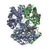
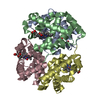
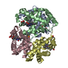
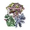
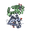
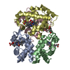
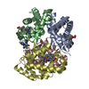
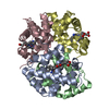
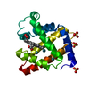
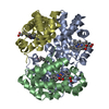
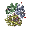
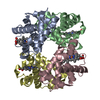
- PDB-2w72: DEOXYGENATED STRUCTURE OF A DISTAL SITE HEMOGLOBIN MUTANT PLUS XE -
+ Open data

- Basic information
| Entry | Database: PDB / ID: 2w72 | ||||||
|---|---|---|---|---|---|---|---|
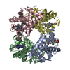
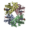
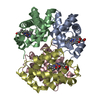
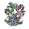
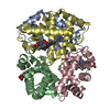
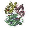
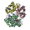
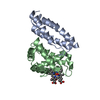
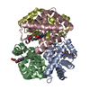
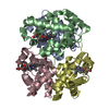
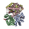
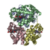
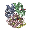
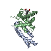
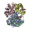
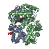
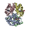
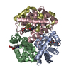
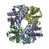
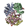
| Title | DEOXYGENATED STRUCTURE OF A DISTAL SITE HEMOGLOBIN MUTANT PLUS XE | ||||||
 Components Components | (HUMAN HEMOGLOBIN ... ) x 3 ) x 3 | ||||||
 Keywords Keywords | OXYGEN TRANSPORT / IRON / HEME / GLYCATION / TRANSPORT / ACETYLATION / PHOSPHOPROTEIN / PACKING DEFECTS / DISEASE MUTATION / DISTAL SITE POINT MUTATION / HYDROPHOBIC CAVITIES / POLYMORPHISM / GLYCOPROTEIN / METAL-BINDING | ||||||
| Function / homology |  Function and homology information Function and homology informationnitric oxide transport / hemoglobin alpha binding / haptoglobin-hemoglobin complex / organic acid binding / cellular oxidant detoxification / hemoglobin binding / renal absorption / hemoglobin complex / oxygen transport / Scavenging of heme from plasma ...nitric oxide transport / hemoglobin alpha binding / haptoglobin-hemoglobin complex / organic acid binding / cellular oxidant detoxification / hemoglobin binding / renal absorption / hemoglobin complex / oxygen transport / Scavenging of heme from plasma / endocytic vesicle lumen / blood vessel diameter maintenance / hydrogen peroxide catabolic process / oxygen carrier activity / Late endosomal microautophagy / Heme signaling / carbon dioxide transport / response to hydrogen peroxide / Erythrocytes take up oxygen and release carbon dioxide / Erythrocytes take up carbon dioxide and release oxygen / Cytoprotection by HMOX1 / platelet aggregation / oxygen binding / regulation of blood pressure / Chaperone Mediated Autophagy / positive regulation of nitric oxide biosynthetic process / tertiary granule lumen / Factors involved in megakaryocyte development and platelet production / blood microparticle / ficolin-1-rich granule lumen / iron ion binding / heme binding / Neutrophil degranulation / extracellular space / extracellular exosome / extracellular region / membrane / metal ion binding / cytosolSimilarity search - Function | ||||||
| Biological species |  HOMO SAPIENS (human) HOMO SAPIENS (human) | ||||||
| Method | X-RAY DIFFRACTION / SYNCHROTRON / MOLECULAR REPLACEMENT / Resolution: 1.07 Å | ||||||
 Authors Authors | Miele, A.E. / Draghi, F. / Sciara, G. / Johnson, K.A. / Renzi, F. / Vallone, B. / Brunori, M. / Savino, C. | ||||||
 Citation Citation | Journal: Biopolymers / Year: 2009 Title: Pattern of Cavities in Globins: The Case of Human Hemoglobin. Authors: Savino, C. / Miele, A.E. / Draghi, F. / Johnson, K.A. / Sciara, G. / Brunori, M. / Vallone, B. | ||||||
| History |
|
- Structure visualization
| Structure viewer | Molecule: MolmilJmol/JSmol |
|---|
- Downloads & links
-Download
| PDBx/mmCIF format | 2w72.cif.gz | 311.8 KB | Display | PDBx/mmCIF format |
|---|---|---|---|---|
| PDB format | pdb2w72.ent.gz | 258 KB | Display | PDB format |
| PDBx/mmJSON format | 2w72.json.gz | Tree view | PDBx/mmJSON format | |
| Others |  Other downloads Other downloads |
-Validation report
| Arichive directory | https://data.pdbj.org/pub/pdb/validation_reports/w7/2w72ftp://data.pdbj.org/pub/pdb/validation_reports/w7/2w72 | HTTPS FTP |
|---|
-Related structure data
| Related structure data |  2w6vC 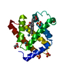 2w6wC  1qi8S S: Starting model for refinement C: citing same article ( |
|---|---|
| Similar structure data |


-Links
 PDBj
PDBj


- Assembly
| Deposited unit | 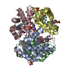
| ||||||||
|---|---|---|---|---|---|---|---|---|---|
| 1 |
| ||||||||
| Unit cell |
|
-Components
-HUMAN HEMOGLOBIN ... , 3 types, 4 molecules ABDC
| #1: Protein | Mass: 15190.353 Da / Num. of mol.: 1 / Fragment: CHAIN ALPHA, RESIDUES 2-142 / Mutation: YES Source method: isolated from a genetically manipulated source Source: (gene. exp.) HOMO SAPIENS (human) / Tissue: BLOOD / Cell: RED BLOOD CELL / Plasmid: PKK223-3 / Production host:  ESCHERICHIA COLI (E. coli) / Strain (production host): TB-1 / References: UniProt: P69905 ESCHERICHIA COLI (E. coli) / Strain (production host): TB-1 / References: UniProt: P69905 | ||
|---|---|---|---|
| #2: Protein | Mass: 15962.263 Da / Num. of mol.: 2 / Fragment: CHAIN BETA, RESIDUES 2-147 / Mutation: YES Source method: isolated from a genetically manipulated source Source: (gene. exp.) HOMO SAPIENS (human) / Tissue: BLOOD / Cell: RED BLOOD CELL / Plasmid: PKK223-3 / Production host: ESCHERICHIA COLI (E. coli) / Strain (production host): TB-1 / References: UniProt: P68871#3: Protein | | Mass: 15222.417 Da / Num. of mol.: 1 / Fragment: CHAIN ALPHA, RESIDUES 2-142 / Mutation: YES Source method: isolated from a genetically manipulated source Source: (gene. exp.) HOMO SAPIENS (human) / Tissue: BLOOD / Cell: RED BLOOD CELL / Plasmid: PKK223-3 / Production host: ESCHERICHIA COLI (E. coli) / Strain (production host): TB-1 / References: UniProt: P69905 |
-Non-polymers , 6 types, 845 molecules 


| #4: Chemical | ChemComp-HEM / Heme B Mass: 616.487 Da / Num. of mol.: 4 / Source method: obtained synthetically / Formula: C34H32FeN4O4 Mass: 616.487 Da / Num. of mol.: 4 / Source method: obtained synthetically / Formula: C34H32FeN4O4#5: Chemical | ChemComp-SO4 / Sulfate Mass: 96.063 Da / Num. of mol.: 8 / Source method: obtained synthetically / Formula: SO4 Mass: 96.063 Da / Num. of mol.: 8 / Source method: obtained synthetically / Formula: SO4#6: Chemical | ChemComp-XE / Xenon Mass: 131.293 Da / Num. of mol.: 15 / Source method: obtained synthetically / Formula: Xe Mass: 131.293 Da / Num. of mol.: 15 / Source method: obtained synthetically / Formula: Xe#7: Chemical |  Mass: 39.098 Da / Num. of mol.: 3 / Source method: obtained synthetically / Formula: K Mass: 39.098 Da / Num. of mol.: 3 / Source method: obtained synthetically / Formula: K#8: Chemical | ChemComp-PO4 / | Phosphate Mass: 94.971 Da / Num. of mol.: 1 / Source method: obtained synthetically / Formula: PO4 Mass: 94.971 Da / Num. of mol.: 1 / Source method: obtained synthetically / Formula: PO4#9: Water | ChemComp-HOH / | WaterMass: 18.015 Da / Num. of mol.: 814 / Source method: isolated from a natural source / Formula: H2O |
|---|
-Details
| Compound details | ENGINEERED RESIDUE IN CHAIN A, LEU 30 TO TYR ENGINEERED RESIDUE IN CHAIN A, HIS 59 TO GLN ...ENGINEERED |
|---|
-Experimental details
-Experiment
| Experiment | Method: X-RAY DIFFRACTION / Number of used crystals: 1 |
|---|
- Sample preparation
| Crystal | Density Matthews: 2.4 Å3/Da / Density % sol: 32.5 % / Description: NONE |
|---|---|
| Crystal grow | pH: 6.5 Details: 3.2M AMMONIUM SULPHATE 0.1M AMMONIUM PHOSPHATE 0.3M K PHOSPHATE PH 6.7 |
-Data collection
| Diffraction | Mean temperature: 100 K |
|---|---|
| Diffraction source | Source: SYNCHROTRON / Site: EMBL/DESY, HAMBURG  / Beamline: BW7A / Wavelength: 0.8453 / Beamline: BW7A / Wavelength: 0.8453 |
| Detector | Type: MARRESEARCH / Detector: CCD / Date: May 9, 2000 |
| Radiation | Protocol: SINGLE WAVELENGTH / Monochromatic (M) / Laue (L): M / Scattering type: x-ray |
| Radiation wavelength | Wavelength: 0.8453 Å / Relative weight: 1 |
| Reflection | Resolution: 1.07→17.6 Å / Num. obs: 250223 / % possible obs: 91.1 % / Observed criterion σ(I): 2 / Redundancy: 4 % / Biso Wilson estimate: 8.54 Å2 / Rmerge(I) obs: 0.04 / Net I/σ(I): 25.8 |
| Reflection shell | Resolution: 1.07→1.12 Å / Redundancy: 3.9 % / Rmerge(I) obs: 0.18 / Mean I/σ(I) obs: 2.5 / % possible all: 90.2 |
- Processing
| Software |
| ||||||||||||||||||||||||||||||||||||||||||||||||||||||||||||||||||||||||||||||||||||||||||||||||||||||||||||||||||||||||||||||||||||||||||||||||||||||||||||||||||||||||||||||||||||||
|---|---|---|---|---|---|---|---|---|---|---|---|---|---|---|---|---|---|---|---|---|---|---|---|---|---|---|---|---|---|---|---|---|---|---|---|---|---|---|---|---|---|---|---|---|---|---|---|---|---|---|---|---|---|---|---|---|---|---|---|---|---|---|---|---|---|---|---|---|---|---|---|---|---|---|---|---|---|---|---|---|---|---|---|---|---|---|---|---|---|---|---|---|---|---|---|---|---|---|---|---|---|---|---|---|---|---|---|---|---|---|---|---|---|---|---|---|---|---|---|---|---|---|---|---|---|---|---|---|---|---|---|---|---|---|---|---|---|---|---|---|---|---|---|---|---|---|---|---|---|---|---|---|---|---|---|---|---|---|---|---|---|---|---|---|---|---|---|---|---|---|---|---|---|---|---|---|---|---|---|---|---|---|---|
| Refinement | Method to determine structure: MOLECULAR REPLACEMENT Starting model: PDB ENTRY 1QI8 Resolution: 1.07→17.32 Å / Cor.coef. Fo:Fc: 0.98 / Cor.coef. Fo:Fc free: 0.974 / SU B: 0.802 / SU ML: 0.019 / Cross valid method: THROUGHOUT / ESU R: 0.029 / ESU R Free: 0.03 / Stereochemistry target values: MAXIMUM LIKELIHOOD / Details: HYDROGENS HAVE BEEN ADDED IN THE RIDING POSITIONS
| ||||||||||||||||||||||||||||||||||||||||||||||||||||||||||||||||||||||||||||||||||||||||||||||||||||||||||||||||||||||||||||||||||||||||||||||||||||||||||||||||||||||||||||||||||||||
| Solvent computation | Ion probe radii: 0.8 Å / Shrinkage radii: 0.8 Å / VDW probe radii: 1.2 Å / Solvent model: MASK | ||||||||||||||||||||||||||||||||||||||||||||||||||||||||||||||||||||||||||||||||||||||||||||||||||||||||||||||||||||||||||||||||||||||||||||||||||||||||||||||||||||||||||||||||||||||
| Displacement parameters | Biso mean: 14.82 Å2
| ||||||||||||||||||||||||||||||||||||||||||||||||||||||||||||||||||||||||||||||||||||||||||||||||||||||||||||||||||||||||||||||||||||||||||||||||||||||||||||||||||||||||||||||||||||||
| Refinement step | Cycle: LAST / Resolution: 1.07→17.32 Å
| ||||||||||||||||||||||||||||||||||||||||||||||||||||||||||||||||||||||||||||||||||||||||||||||||||||||||||||||||||||||||||||||||||||||||||||||||||||||||||||||||||||||||||||||||||||||
| Refine LS restraints |
|